When I woke up and my mind and spirit were reborn awaking from the brainwashing that I was raised to believe as a child and as a young man in a sports driven and Hollywood propaganda based mainstream media society. My family and friends rejected me my treatment was that of a black sheep and outcast from the Matrix that they were programmed to believe in with fake News, science, history and religion based system. I began to write blogs and find like minded friends who began to be my tribe and now is a army of truth seeking Patriots!
Now day’s our work consists of being a watchdog group and doing community services such as courtroom observation though a organization that I am a cofounder of along with my good friend Eric Jones nicknamed the Freedom Screamer of CourtroomWatch.com to find out more information about this topic please visit the website because it’s just to much to describe exactly what we do but basically we are advocating for justice and freedom for all.
As far as being a watchdog group we are an independent news source. Because of my connections and networks around the world we often get information that the main stream media is not privy to. Our sources come from our personal network of activists and grassroots sources who are boots on the ground. We are a grassroots political organization dedicated to Truth Justice and freedom not just for America for the entire world.
Early on before the anti-DeepState Party became a global community I was an advocate against child trafficking and have been contacted by several families over the years to try and tell their stories to get the word out about such things happening right in my own home state of NY.
Also we have been an advocate for health and wellness we are part of the anti-vaccination movement. This was definitely part of the reason that my family treated me like a black sheep outcast. I would often warn them about agenda 21 eugenics in the global plan for depopulation using a bio weapon through vaccines.
My first real eye-opener to the pharmaceutical industry was with a interview that I did with a group of guys on the radio station 1310 AM out of Brockport New York we talk to a doctor Charles Pixley who was using holistic remedies to cure cancer called 714X. Dr. Charles Pytchley was arrested and jailed silenced. He was told by the courts that he could never write a book or publicize his findings because 714X Was not an approved drug by the FDA. Even though during the time that he was using this it was not considered a synthetic pharmaceutical drug it was a naturalholistic remedy.
But what we’re doing nowadays is building a coalition a community of Activists Fighting government corruption, big tech censorship. Our Independent news media website studio 1776 has been completely scrubbed or censored from main stream social media and I’m afraid that not long from now even this website will be totally blocked deactivated from main stream social media because of our advocacy and being a watchdog group for conservatives values.
Oklahoma State Senator Ralph Shortey introduced a bill that would ban “the manufacture or sale of food or products which use aborted human fetuses.”
PepsiCo for working with a company called Senomyx that “has been accused of using proteins derived from human embryonic kidney cells in its research.” Address San Diego, CA 92121-3051
United States Phone+1-858-6468300 Fax 858-4040752 Webwww.senomyx.com
HEK 293 cells But some food companies are using cell lines that were originally derived from human fetuses in order to develop new food products. … The cells, called HEK 293 cells (that stands for human embryonic kidney) were taken from an aborted fetus in the 1970s in the Netherlands. Human cloned DNA in your foods people no joke
The cells, called HEK 293 cells (that stands for human embryonic kidney) were taken from an aborted fetus in the 1970s in the Netherlands.
The Original Unnamed aborted babies cell culture cell line isn’t leading to new abortions but it sure is human DNA.
Origins of the HEK293 Cell Line
HEK293 is a cell line derived from human embryonic kidney cells grown in tissue culture. They are also known, more informally, as HEK cells. This particular line was initiated by the transformation and culturing of normal HEK cells with sheared adenovirus 5 DNA. The transformation resulted in the incorporation of approximately 4.5 kilobases from the viral genome into human chromosome 19 of the HEK cells. The line was cultured by scientist Alex Van der Eb in the early 1970s at his lab at the University of Leiden, Holland. The transformation was executed by Frank Graham, another scientist Van der Eb’s lab who invented the calcium phosphate method for transfecting cells. The source of the cells was a healthy aborted fetus of unknown parenthood. The name HEK293 is thusly named because it was Frank Graham’s 293rd experiment.
The type of kidney cell that the HEK293 cell line came arose from is unknown and it is difficult to conclusively characterize the cells post-transformation since adenovirus 5 could have significantly disrupted cell morphology and expression. Also, embryonic kidneys are a heterogeneous mix of almost all the types of cells present in the body. In fact, it has been speculated by independent researchers, including Van der Eb himself, that the cells may be neuronal in origin. Although theoretically possible, most cells derived from an embryonic kidney would be endothelial, epithelial or fibroblast cells. Neuronal origin is suspected due to the presence of mRNA and gene products typically found in neurons.
Today, HEK293 cells are frequently used in cell biology and biotechnology, second only to HeLa, the first human cell line. Around establishment of HeLa in 1951, scientists were reluctant to accept and use human cell lines out of concern for an oncogenic agent in them. This concern, along with the known ability of animal cell lines to grow rapidly and yield a high amount of proteins, gave scientists reason to favor animal cell lines over human cell lines when producing recombinant proteins. However, advances in technology since then have allowed for an increase in human cell line use. One advantage of human cell lines is that they are able to produce proteins most similar to those that humans naturally synthesize. Now there are approved recombinant biotherapeutic products produced from HEK293 and other human cell lines.
HEK293 and its derivatives are used in a wide range of experiments, including signal transduction and protein interaction studies, rapid small-scale protein production, and biopharmaceutical production. HEK293 cells easily grow in suspension serum-free culture, reproduce rapidly, and produce high levels of protein, which explains why they have been widely used to produce research-grade proteins for a number of years.
“We’re helping companies clean up their labels,” said Senomyx’s chief executive, Kent Snyder.
Senomyx, based in San Diego, uses many of the same research techniques that biotechnology companies apply in devising new drugs. Executives say that a taste receptor or family of receptors on the tongue or in the mouth are responsible for recognizing a taste. Using the human genome sequence, the company says, it has identified hundreds of those taste receptors. Its chemical compounds activate the receptors in a way that accentuates the taste of sugar or salt. It is still experimenting to determine the most potent compounds, its chief scientist, Mark Zoller, said.
But Senomyx maintains that its new products are safe because they will be used in tiny quantities.
Kraft, Nestlé, Coca-Cola and Campbell Soup have contracted with Senomyx for exclusive rights to use the ingredients in certain types of food and beverages, although the companies declined to identify those categories.
Elise Wang, an analyst at Smith Barney, said that Kraft was planning to use Senomyx’s sweet flavoring to reduce the sugar in powdered beverages like Kool-Aid by one-third. Campbell Soup, she said, is looking at cutting sodium levels by a third with the salt flavoring.
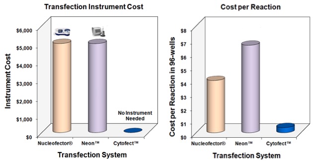
Cost Effective
Since Cytofect™ Transfection Reagents require no expensive instruments to use, high efficiency transfection of primary cells becomes much less costly.
With no upfront investment in electroporation devices, your lab can save its precious research budget to focus on downstream assays instead.
Senomyx is an Americanbiotechnology company working toward developing additives to amplify certain flavors and smells in foods. The company claims to have essentially “reverse engineered” the receptors in humans that react for taste and aroma, and that they are capitalizing on these discoveries to produce chemicals that will make food taste better. On 17 Sept 2018, Firmenich completed the acquisition of Senomyx. [1]
Senomyx develops patented flavor enhancers by using “proprietary taste receptor-based assay systems”, which have been previously expressed in human cell culture, in HEK293 cells.[2]
HEK293 cells are a cell line widely used in biological and medical research, immortalised through a genetic modification removed from the original human embryonic kidney cells taken from a healthy, electively aborted human fetus in the early 1970s.[3] The receptors in the assay are used to identify flavours; they are not used as flavours themselves. No human taste receptors are used as ingredients in any flavourings. Using information from the human genome sequence, Senomyx has identified hundreds of taste receptors and currently owns 113 patents on their discoveries. Senomyx collaborates with seven of the world’s largest food companies to further their research and to fund development of their technology.
Cell Applications, Inc
5820 Oberlin Drive, Suite 101
San Diego, CA 92121
Open M-F, 8am-5pm PST
types, complimented by optimized products to serve life science R&D … media like DMEM and RPMI, and immortalized cell lines (HeLa, HEK 293 … testing and certification, so the procedures and product remains consistent. …
I. TERMS OF USE Cell Applications, Inc. (“CAI”) will sell products … of the CAI’s products (such as to collect a debt, resolve a dispute … by delays in receiving orders. VI. PRODUCT USE AND RECOMMENDATIONS All …
Srihirun, S., Park, J. W., Teng, R., Sawaengdee, W., Piknova, B., & Schechter, A. N. (2019). Nitrate uptake and metabolism in human skeletal muscle cell cultures. Nitric Oxide.
Human Aortic Smooth Muscle Cells: HAOSMC
Lu, Y., Sun, X., Peng, L., Jiang, W., Li, W., Yuan, H., & Cai, J. (2019). Angiotensin II-Induced vascular remodeling and hypertension involves cathepsin L/V-MEK/ERK mediated mechanism. International Journal of Cardiology.
Human Dermal Fibroblasts: HDF
Chaudhuri, R. K., Meyer, T., Premi, S., & Brash, D. Acetyl Zingerone (2019): An efficacious multifunctional ingredient for continued protection against on‐going DNA damage in melanocytes after sun exposure ends. International Journal of Cosmetic Science.
Human Coronary Artery Endothelial Cells: HCAEC
Gamon, L. F., Dieterich, S., Ignasiak, M. T., Schrameyer, V., & Davies, M. J. (2019). Iodide modulates protein damage induced by the inflammation-associated heme enzyme myeloperoxidase. Redox Biology, 101331.
Rat Dermal Fibroblasts: RDF
Palungwachira, P., Tancharoen, S., Phruksaniyom, C., Klungsaeng, S., Srichan, R., Kikuchi, K., & Nararatwanchai, T. (2019). Antioxidant and Anti-Inflammatory Properties of Anthocyanins Extracted from Oryza sativa L. in Primary Dermal Fibroblasts. Oxidative Medicine and Cellular Longevity, 2019.
Human Dermal Fibroblasts: HDF
Wang, X., Hong, H., & Wu, J. (2019). Hen collagen hydrolysate alleviates UVA-induced damage in human dermal fibroblasts. Journal of Functional Foods, 63, 103574.
Human Epidermal Keratinocytes: HEK
Chaudhuri, R. K., Meyer, T., Premi, S., & Brash, D. Acetyl Zingerone (2019): An efficacious multifunctional ingredient for continued protection against on‐going DNA damage in melanocytes after sun exposure ends. International Journal of Cosmetic Science.
Human Umbilical Vein Endothelial Cells: HUVEC
Matsunuma, S., Handa, S., Kamei, D., Yamamoto, H., Okuyama, K., & Kato, Y. (2019). Oxaliplatin induces prostaglandin E2 release in vascular endothelial cells. Cancer Chemotherapy and Pharmacology, 1-6.
Human Pulmonary Artery Endothelial Cells: HPAEC
Blais-Lecours, P., Laouafa, S., Arias-Reyes, C., Santos, W. L., Joseph, V., Burgess, J. K., … & Marsolais, D. (2019). Metabolic adaptation of airway smooth muscle cells to a SPHK2 substrate precedes cytostasis. American Journal of Respiratory Cell and Molecular Biology,
Bovine Aortic Endothelial Cells: BAOEC
Ogata, F., Nakamura, T., Nakajima, M., Toda, M., Otani, M., & Kawasaki, N. (2019). PO43− adsorption in a complex solution by nickel–cobalt hydroxide, and its cytotoxicity on bovine aortic endothelial cells. Journal of Environmental Chemical Engineering.
MesoEndo Cell Growth Medium
Detsika, M. G., Myrtsi, E. D., Koulocheri, S. D., Haroutounian, S. A., Lianos, E. A., & Roussos, C. (2019). Induction of decay accelerating factor and membrane cofactor protein by resveratrol attenuates complement deposition in human coronary artery endothelial cells. Biochemistry and Biophysics Reports, 19, 100652.
Rat Pulmonary Artery Smooth Muscle Cells: RPASMC
Suzuki, Y. J., Marcocci, L., Shimomura, T., Tatenaka, Y., Ohuchi, Y., & Brelidze, T. I. (2019). Protein Redox State Monitoring Studies of Thiol Reactivity. Antioxidants, 8 (5), 143.
Human Coronary Artery Endothelial Cells: HCAEC
Lorentzen, L. G., Chuang, C. Y., Rogowska-Wrzesinska, A., & Davies, M. J. (2019). Identification and quantification of sites of nitration and oxidation in the key matrix protein laminin and the structural consequences of these modifications. Redox Biology, 101226.
Human Liver, Spleen, Kidney and Testes RNA
Swystun, L. L., Ogiwara, K., Lai, J. D., Ojala, J. R., Rawley, O., Lassalle, F., … & Tryggvason, K. (2019). The scavenger receptor SCARA 5 is an endocytic receptor for von Willebrand factor expressed by littoral cells in the human spleen. Journal of Thrombosis and Haemostasis.
Human Umbilical Vein Endothelial Cells: HUVEC
Brines, M. and Cerami, A., (2019). TISSUE PROTECTIVE PEPTIDES FOR PREVENTING AND TREATING DISEASES AND DISORDERS ASSOCIATED WITH TISSUE DAMAGE. U.S. Patent Application 16/096,247.
Human Dermal Fibroblasts: HDF
Yang, H., Sun, J., Chen, H., Wang, F., Li, Y., Wang, H., & Qu, T. (2019). Mesenchymal stem cells from bone marrow attenuated the chronic morphine-induced cAMP accumulation in vitro. Neuroscience letters, 698, 76-80.
Human EpiVita Serum-Free Growth Medium
Lin, E. S., Chang, W. A., Chen, Y. Y., Wu, L. Y., Chen, Y. J., & Kuo, P. L. (2019). Deduction of Novel Genes Potentially Involved in Keratinocytes of Type 2 Diabetes Using Next-Generation Sequencing and Bioinformatics Approaches. Journal of clinical medicine, 8(1), 73.
Human Carotid Artery Smooth Muscle Cells: HCtASMC
Aldi, S., Eriksson, L., Kronqvist, M., Lengquist, M., Löfling, M., Folkersen, L…& Österholm, C. (2019). Dual roles of heparanase in human carotid plaque calcification. Atherosclerosis.
Human Umbilical Vein Endothelial Cells: HUVEC
Swaminathan, S., Hamid, Q., Sun, W., & Clyne, A. M. (2019). Bioprinting of 3D breast epithelial spheroids for human cancer models. Biofabrication.
MesoEndo Cell Growth Medium
Pott, G. B., Tsurudome, M., Proctor, L. L., & Goalstone, M. L. (2019). CIGARETTE SMOKE EXTRACT, KALLIKREIN-6 AND APROTININ REGULATE PRODUCTION OF SOLUBLE VCAM-1 AND ICAM-1 IN HUMAN CAROTID ENDOTHELIAL CELLS.
Human Epidermal Keratinocytes: HEK
Yamakami, Y., Morino, K., Takauji, Y., Kasukabe, R., Miki, K., Hossain, M. N., … & Fujii, M. (2019). Extract of Emblica officinalis enhances the growth of human keratinocytes in culture. Journal of integrative medicine.
Human Bladder Epithelial Cells: HBlEpC
Kim, D., Ahn, B. N., Kim, Y., Hur, D. Y., Yang, J. W., Park, G. B., … & Kim, M. K. (2019). High Glucose with Insulin Induces Cell Cycle Progression and Activation of Oncogenic Signaling of Bladder Epithelial Cells Cotreated with Metformin and Pioglitazone. Journal of diabetes research, 2019.
Human Carotid Artery Endothelial Cells: HCtAEC
Pott, G. B., Tsurudome, M., Proctor, L. L., & Goalstone, M. L. (2019). CIGARETTE SMOKE EXTRACT, KALLIKREIN-6 AND APROTININ REGULATE PRODUCTION OF SOLUBLE VCAM-1 AND ICAM-1 IN HUMAN CAROTID ENDOTHELIAL CELLS.
Human Dermal Fibroblasts: HDF
Desai, D., Lauver, M. D., Cruz, L., Jin, G., Ferguson, K., Roper, B., … & Buchkovich, N. J. (2019). Inhibition of Diverse Opportunistic Viruses by Structurally Optimized Retrograde Trafficking Inhibitors. Bioorganic & Medicinal Chemistry.
Human Mammary Epithelial Cells: HMEpC
Fukui, T., Soda, K., Takao, K., & Rikiyama, T. (2019). Extracellular spermine activates DNA methyltransferase 3A and 3B. International journal of molecular sciences, 20(5), 1254.
Rat Aortic Endothelial Cells: RAOEC
Naik, J. S., & Walker, B. R. (2018). Endothelial-dependent dilation following chronic hypoxia involves TRPV4-mediated activation of endothelial BK channels. Pflügers Archiv-European Journal of Physiology, 470(4), 633-648.
2018
Human Chondrocytes
Chen, Y.J., Chang, W.A., Wu, L.Y., Hsu, Y.L., Chen, C.H. and Kuo, P.L., 2018. Systematic Analysis of Transcriptomic Profile of Chondrocytes in Osteoarthritic Knee Using Next-Generation Sequencing and Bioinformatics. Journal of Clinical Medicine, 7(12), p.535.
Bovine Aortic Endothelial Cells: BAOEC
Takahashi, A., Takahashi, M., Fujie, T., Hara, T., Yoshida, E., Yamamoto, C. and Kaji, T., 2018. A zinc complex that suppresses the expression of a reactive sulfur species-producing enzyme, cystathionine γ-lyase, in cultured vascular endothelial cells. Fundamental Toxicological Sciences, 5(6), pp.181-184.
Human Dermal Fibroblasts: HDF
Yu, C., Ma, X., Zhu, W., Wang, P., Miller, K.L., Stupin, J., Koroleva-Maharajh, A., Hairabedian, A. and Chen, S., 2018. Scanningless and continuous 3D bioprinting of human tissues with decellularized extracellular matrix. Biomaterials.
Human Umbilical Vein Endothelial Cells: HUVEC
Tan, Z. B., Fan, H. J., Wu, Y. T., Xie, L. P., Bi, Y. M., Xu, H. L., … & Zhou, Y. C. (2018). Rheum palmatum extract exerts anti-hepatocellular carcinoma effects by inhibiting signal transducer and activator of transcription 3 signaling. Journal of Ethnopharmacology.
Skeletal Muscle Growth Medium
Patton, J. B., Bennuru, S., Eberhard, M. L., Hess, J. A., Torigian, A., Lustigman, S., … & Abraham, D. (2018). Development of Onchocerca volvulus in humanized NSG mice and detection of parasite biomarkers in urine and serum. PLOS Neglected Tropical Diseases, 12(12), e0006977.
Human Chondrocytes
Tsumaki, N. and Yamashita, A., Kyoto University, 2018. Prophylactic and therapeutic agents for fgfr3 diseases and screening method for the same. U.S. Patent Application 16/059,462.
Human Dermal Fibroblasts: HDF
Playne, R., Jones, K. S., & Connor, B. (2018). Generation of dopamine neuronal-like cells from induced neural precursors derived from adult human cells by non-viral expression of lineage factors. J Stem Cells Regen Med.
Human Dermal Fibroblasts: HDF
Ikeda, K., Uchida, N., Nishimura, T., White, J., Martin, R.M., Nakauchi, H., Sebastiano, V., Weinberg, K.I. and Porteus, M.H., (2018). Efficient scarless genome editing in human pluripotent stem cells. Nature methods, 15(12), p.1045.
Endothelial Cell Growth Medium Leonard, J.N., Stranford, D.M. and Passineau, M.J., Northwestern University, (2018). Deliverable extracellular vesicles incorporating cell membrane transport proteins. U.S. Patent Application 15/975,222.
Human Peripheral Blood B Cells: HPBB
Marin, E.H., Paek, H., Li, M., Ban, Y., Karaga, M.K., Shashidharamurthy, R. and Wang, X., 2018. Caffeic acid phenethyl ester exerts apoptotic and oxidative stress on human multiple myeloma cells. Investigational new drugs, pp.1-12.
Human Adipocyte Differentiation Medium
Bagher, Z., Kamrava, S. K., Alizadeh, R., Farhadi, M., Absalan, M., Falah, M. & Komeili, A. (2018). Differentiation of Neural Crest Stem Cells From Nasal Mucosa into Motor Neuron-Like Cells. Journal of Chemical Neuroanatomy.
MCDB 105 Medium
Starbuck, K., Al-Alem, L., Eavarone, D. A., Hernandez, S. F., Bellio, C., Prendergast, J. M., & Behrens, J. (2018). Treatment of ovarian cancer by targeting the tumor stem cell-associated carbohydrate antigen, Sialyl-Thomsen-nouveau. Oncotarget, 9(33), 23289.
Bovine Pulmonary Artery Endothelial cells: BPAEC
Rowan, S. C., Rochfort, K. D., Piouceau, L., Cummins, P. M., O’Rourke, M., & McLoughlin, P. (2018). Pulmonary endothelial permeability and tissue fluid balance depend on the viscosity of the perfusion solution. American Journal of Physiology-Lung Cellular and Molecular Physiology.
Human Dermal Fibroblasts: HDF
Chaudhuri, R.K., Sytheon Ltd, 2018. Skin enhancing compositions and methods. U.S. Patent Application 15/798,804.
Human Preadipocytes: HPAd
Matsubara, Yumiko, Takeru Zama, Yasuo Ikeda, Yukako Uruga, Toshio Suda, and Sahoko Matsuoka. “Method for producing megakaryocytes, platelets and/or thrombopoietin using mesenchymal cells.” U.S. Patent Application 15/815,069.
Human Aortic Smooth Muscle Cells: HAOSMC
van Engeland, N. C., Pollet, A. M., den Toonder, J. M., Bouten, C. V., Stassen, O. M., & Sahlgren, C. M. (2018). A biomimetic microfluidic model to study signalling between endothelial and vascular smooth muscle cells under hemodynamic conditions. Lab on a Chip.
Canine Osteoblasts: CnOb
Scott, M.C., Sarver, A.L., Modiano, J.F., Subramanian, S., Largaespada, D.A. and Spector, L.G., University of Minnesota, 2018. Tumor Analytical Methods. U.S. Patent Application 15/783,352.
Human Dermal Fibroblasts: HDF
Yoshida, Shunsuke, Mitsuru Inamura, Tohru Tanaka, Hiroyuki Ishikawa, and Hidenori Ito. “Stem cell removing method, differentiated cell protective method, and culture medium composition.” U.S. Patent Application 15/565,422.
Human Chondrocytes: HC
Li, A., Wei, Y., Hung, C., & Vunjak-Novakovic, G. (2018). Chondrogenic properties of collagen type XI, a component of cartilage extracellular matrix. Biomaterials.
Human Coronary Artery Endothelial Cells: HCAEC
Xu, S., Xu, Y., Yin, M., Zhang, S., Liu, P., Koroleva, M.,..& Jin, Z. G. (2018). Flow-dependent epigenetic regulation of IGFBP5 expression by H3K27me3 contributes to endothelial anti-inflammatory effects. Theranostics, 8(11), 3007-3021.
Human MesoEndo Endothelial Cell Media
Xu, S., Xu, Y., Yin, M., Zhang, S., Liu, P., Koroleva, M.,..& Jin, Z. G. (2018). Flow-dependent epigenetic regulation of IGFBP5 expression by H3K27me3 contributes to endothelial anti-inflammatory effects. Theranostics, 8(11), 3007-3021.
Rat Aortic Smooth Muscle Cells: RAOSMC
Park, H. S., Han, J. H., Jung, S. H., Lee, D. H., Heo, K. S., & Myung, C. S. (2018). Anti-apoptotic effects of autophagy via ROS regulation in microtubule-targeted and PDGF-stimulated vascular smooth muscle cells. The Korean Journal of Physiology & Pharmacology, 22(3), 349-360.
Human Dermal Fibroblasts: HDF
Kikkawa, Y., Enomoto-Okawa, Y., Fujiyama, A., Fukuhara, T., Harashima, N., Sugawara, Y., … & Ito, Y. (2018). Internalization of CD239 highly expressed in breast cancer cells: a potential antigen for antibody-drug conjugates. Scientific reports, 8.
Human Pulmonary Artery Smooth Muscle Cells: HPASMC
Wilson, J. L., Warburton, R., Taylor, L., Toksoz, D., Hill, N., & Polgar, P. (2018). Unraveling endothelin-1 induced hypercontractility of human pulmonary artery smooth muscle cells from patients with pulmonary arterial hypertension. PloS one, 13(4), e0195780.
Human Dermal Fibroblasts: HDF
Ito, Tomohisa, Takashi Ando, Miki Suzuki-Karasaki, Tomohiko Tokunaga, Yukihiro Yoshida, Toyoko Ochiai, Yasuaki Tokuhashi, and Yoshihiro Suzuki-Karasaki. “Cold PSM, but not TRAIL, triggers autophagic cell death: A therapeutic advantage of PSM over TRAIL.” International Journal of Oncology.
Human Carotid Artery Endothelial Cells: HCtAEC
Hoh, B. L., Rojas, K., Lin, L., Fazal, H. Z., Hourani, S., Nowicki, K. W., … & Hosaka, K. (2018). Estrogen Deficiency Promotes Cerebral Aneurysm Rupture by Upregulation of Th17 Cells and Interleukin‐17A Which Downregulates E‐Cadherin. Journal of the American Heart Association, 7(8), e008863.
Sakima, M., Hayashi, H., Al Mamun, A., & Sato, M. (2018). VEGFR-3 signaling is regulated by a G-protein activator, activator of G-protein signaling 8, in lymphatic endothelial cells. Experimental cell research.
Human Dermal Fibroblasts: HDF
Kang, L., Liu, X., Yue, Z., Chen, Z., Baker, C., Winberg, P. C., & Wallace, G. G. (2018). Fabrication and In Vitro Characterization of Electrochemically Compacted Collagen/Sulfated Xylorhamnoglycuronan Matrix for Wound Healing Applications. Polymers, 10(4), 415.
Human Chondrocyte Media
Barrett, Carolyn, and Yaling Shi. “Cartilage mosaic compositions and methods.” U.S. Patent Application 15/608,679.
Human Dermal Fibroblasts: HDF
Esparza, Y., Bandara, N., Ullah, A., & Wu, J. (2018). Hydrogels from feather keratin show higher viscoelastic properties and cell proliferation than those from hair and wool keratins. Materials Science and Engineering: C.
Human Aortic Smooth Muscle Cells: HAOSMC
Cardenas, C. L. L., Kessinger, C. W., Cheng, Y., MacDonald, C., MacGillivray, T., Ghoshhajra, B., … & Kaminski, N. (2018). An HDAC9-MALAT1-BRG1 complex mediates smooth muscle dysfunction in thoracic aortic aneurysm. Nature Communications, 9(1), 1009.
Human Epidermal Keratinocytes: HEK
Takahashi, A., Loo, T. M., Okada, R., Kamachi, F., Watanabe, Y., Wakita, M., & Ohtani, N. (2018). Downregulation of cytoplasmic DNases is implicated in cytoplasmic DNA accumulation and SASP in senescent cells. Nature Communications, 9(1), 1249.
Bovine Aortic Endothelial Cells: BAOEC
Zhao, X., Hui, D. S., Lee, R., & Edwards, J. L. (2018). Ratiometric quantitation of thiol metabolites using non-isotopic mass tags. Analytica Chimica Acta.
Human Endothelial Cell Growth Medium
Passineau, M.J., Murali, S., Benza, R.L. and Pollett, J.B., Allegheny-Singer Research Institute, 2018. ISOLATION OF PULMONARY ARTERIAL ENDOTHELIAL CELLS FROM PATIENTS WITH PULMONARY VASCULAR DISEASE AND USES THEREOF. U.S. Patent Application 15/806,751.
DiI-Ac-LDL Kit
Lian, W., Hu, X., Shi, R., Han, S., Cao, C., Wang, K., & Li, M. (2018). MiR-31 regulates the function of diabetic endothelial progenitor cells by targeting Satb2. Acta biochimica et biophysica Sinica.
Human Hair Follicle Dermal Papilla Cells: HFDPC
Lahiji SF, Seo SH, Kim S, Dangol M, Shim J, Li CG, Ma Y, Lee C, Kang G, Yang H, Choi KY. (2018). Transcutaneous implantation of valproic acid-encapsulated dissolving microneedles induces hair regrowth. Biomaterials.
Bovine Aortic Endothelial Cells: BAOEC
Zhao, X., Hui, D. S., Lee, R., & Edwards, J. L. (2018). Ratiometric quantitation of thiol metabolites using non-isotopic mass tags. Analytica Chimica Acta.
Human Aortic Smooth Muscle Cells: HAOSMC
Cardenas, C. L. L., Kessinger, C. W., MacDonald, C., Jassar, A. S., Isselbacher, E. M., Jaffer, F. A., & Lindsay, M. E. (2018). Inhibition of the methyltranferase EZH2 improves aortic performance in experimental thoracic aortic aneurysm. JCI insight, 3(5).
Endothelial Cell Growth Medium
CD Nichols, B YU (2018). LOW DOSAGE SEROTONIN 5-HT2A RECEPTOR AGONIST TO SUPPRESS INFLAMMATION. US Patent App. 15/478,437.
Rat Brain Microvascular Endothelial Cells: RBMVEC
Brailoiu, E., Barlow, C. L., Ramirez, S. H., Abood, M. E., & Brailoiu, G. C. (2018). Effects of Platelet-Activating Factor on brain microvascular endothelial cells. Neuroscience.
Human Carotid Artery Smooth Muscle Cells: HCtASMC
Han, X., Sakamoto, N., Tomita, N., Meng, H., Sato, M., & Ohta, M. (2017). Influence of shear stress on phenotype and MMP production of smooth muscle cells in a co-culture model. Journal of Biorheology, 31(2), 50-56.
Human Fibroblast-Like Synoviocytes: HFLS
Yu, R., Li, C., Sun, L., Jian, L., Ma, Z., Zhao, J., & Liu, X. (2018). Hypoxia induces production of citrullinated proteins in human fibroblast‐like synoviocytes through regulating HIF1α. Scandinavian journal of immunology.
Human Cardiac Fibroblasts: HCF
John, C.M., Meenakshi, G.A.U.R., Matthew, L. and Wang, X., MANDALMED Inc, 2018. Methods and compositions for preventing and treating damage to the heart. U.S. Patent Application 15/666,456.
Rat Smooth Muscle Cell Media
Chinnappan, M., Mohan, A., Agarwal, S., Dalvi, P., & Dhillon, N. K. (2018). Network of MicroRNAs Mediate Translational Repression of Bone Morphogenetic Protein Receptor‐2: Involvement in HIV‐Associated Pulmonary Vascular Remodeling. Journal of the American Heart Association, 7(5), e008472.
Human Smooth Muscle Cell Growth Medium
Cardenas, C. L. L., Kessinger, C. W., MacDonald, C., Jassar, A. S., Isselbacher, E. M., Jaffer, F. A., & Lindsay, M. E. (2018). Inhibition of the methyltranferase EZH2 improves aortic performance in experimental thoracic aortic aneurysm. JCI insight, 3(5).
Human Fibroblast-Like Synoviocytes: HFLS
Rosa, I., Marini, M., Guasti, D., Ibba-Manneschi, L., & Manetti, M. (2018). Morphological evidence of telocytes in human synovium. Scientific reports, 8(1), 3581.
Human Carotid Artery Endothelial Cells: HCtAEC
Han, X., Sakamoto, N., Tomita, N., Meng, H., Sato, M., & Ohta, M. (2017). Influence of shear stress on phenotype and MMP production of smooth muscle cells in a co-culture model. Journal of Biorheology, 31(2), 50-56.
Human Fibroblast-Like Synoviocytes: Rheumatoid Arthritis: HFLS-RA
Hagihara, M., Shimizu, M. and Wada, Y., Ube Industries Ltd, 2018. Method of producing substance. U.S. Patent Application 15/545,624.
Bovine Aortic Endothelial Cells: BAOEC
Uhl, C. G., Gao, Y., Zhou, S., & Liu, Y. (2018). The shape effect on polymer nanoparticle transport in a blood vessel. RSC Advances, 8(15), 8089-8100.
Human Umbilical Vein Endothelial Cells: HUVEC
Sasahira, T., Nishiguchi, Y., Kurihara-Shimomura, M., Nakashima, C., Kuniyasu, H., & Kirita, T. (2018). NIPA-like domain containing 1 is a novel tumor-promoting factor in oral squamous cell carcinoma. Journal of cancer research and clinical oncology, 1-8.
Human Fibroblast-Like Synoviocytes: Rheumatoid Arthritis: HFLS-RARhys, H. I., Dell’Accio, F., Pitzalis, C., Moore, A., Norling, L. V., & Perretti, M. (2018). Neutrophil Microvesicles from Healthy Control and Rheumatoid Arthritis Patients Prevent the Inflammatory Activation of Macrophages. EBioMedicine.
Rabbit Aortic Smooth Muscle Cells: RbAOSMC
Honda, K., Matoba, T., Antoku, Y., Koga, J. I., Ichi, I., Nakano, K., & Egashira, K. (2018). Lipid-Lowering Therapy With Ezetimibe Decreases Spontaneous Atherothrombotic Occlusions in a Rabbit Model of Plaque ErosionHighlights: A Role of Serum Oxysterols. Arteriosclerosis, thrombosis, and vascular biology, 38(4), 757-771.
Human Dermal Fibroblasts: HDF
Tokunaga, T., Ando, T., Suzuki-Karasaki, M., Ito, T., Onoe-Takahashi, A., Ochiai, T., Soma, M. and Suzuki-Karasaki, Y., 2018. Plasma-stimulated medium kills TRAIL-resistant human malignant cells by promoting caspase-independent cell death via membrane potential and calcium dynamics modulation. International journal of oncology, 52(3), pp.697-708.
Human Coronary Artery Endothelial Cells RNA
Baggio, L. L., Yusta, B., Mulvihill, E. E., Cao, X., Streutker, C. J., Butany, J., & Drucker, D. J. (2018). GLP-1 receptor expression within the human heart. Endocrinology, 159(4), 1570-1584.
Grunlan, M.A., Cote, G.L., Abraham, A.A., Fei, R. and Locke, A.K., Texas A&M University System, 2018. Self-Cleaning Membrane for Medical Devices. U.S. Patent Application 15/545,811.
Bovine Aortic Smooth Muscle Cells: BAOSMC
Tsukagoshi, T., Nguyen, T. V., Shoji, K. H., Takahashi, H., Matsumoto, K., & Shimoyama, I. (2018). Cellular dynamics of bovine aortic smooth muscle cells measured using MEMS force sensors. Journal of Physics D: Applied Physics, 51(14), 145401.
Rat Fibroblast Growth Medium
Grunlan, M.A., Cote, G.L., Abraham, A.A., Fei, R. and Locke, A.K., Texas A&M University System, 2018. Self-Cleaning Membrane for Medical Devices. U.S. Patent Application 15/545,811.
Human Umbilical Vein Endothelial Cells: HUVEC
Gaston, B.M., Straub, A.C., Isakson, B.E. and Columbus, L., University of Virginia Licensing and Ventures Group, 2018. Compositions and methods for regulating arterial tone. U.S. Patent Application 15/643,633.
Rat Aortic Endothelial Cells: RAOEC
Naik, J.S. and Walker, B.R., 2018. Endothelial-dependent dilation following chronic hypoxia involves TRPV4-mediated activation of endothelial BK channels. Pflügers Archiv-European Journal of Physiology, pp.1-16.
Human Fibroblast-Like Synoviocytes: HFLS
Hagihara, M., Shimizu, M. and Wada, Y., Ube Industries Ltd, 2018. Method of producing substance. U.S. Patent Application 15/545,624.
Human Preadipocytes: HPAd
Oishi, T., Sakata, A., Shishido, M. and Hirakawa, S., A serum protein, an unexpected player inducing the skin sagging, and a proposed measure for improving the facial sagging.
Human Adipocyte Differentiation Medium Oishi, T., Sakata, A., Shishido, M. and Hirakawa, S., A serum protein, an unexpected player inducing the skin sagging, and a proposed measure for improving the facial sagging.
Human Umbilical Vein Smooth Muscle Cells: HUVSMC
Gaston, B.M., Straub, A.C., Isakson, B.E. and Columbus, L., University of Virginia Licensing and Ventures Group, 2018. Compositions and methods for regulating arterial tone. U.S. Patent Application 15/643,633.
Rat Aortic Endothelial Cells: RAOEC
Iba, T., Hirota, T., Sato, K. and Nagaoka, I., 2018. Protective effect of a newly developed fucose-deficient recombinant antithrombin against histone-induced endothelial damage. International Journal of Hematology, pp.1-7.
Human Dermal Fibroblasts: HDF
Ito, N., Katoh, K., Kushige, H., Saito, Y., Umemoto, T., Matsuzaki, Y., Kiyonari, H., Kobayashi, D., Soga, M., Era, T. and Araki, N., 2018. Ribosome Incorporation into Somatic Cells Promotes Lineage Transdifferentiation towards Multipotency. Scientific reports, 8(1), p.1634.
Human dermal fibroblast growth medium
Ito, N., Katoh, K., Kushige, H., Saito, Y., Umemoto, T., Matsuzaki, Y., Kiyonari, H., Kobayashi, D., Soga, M., Era, T. and Araki, N., 2018. Ribosome Incorporation into Somatic Cells Promotes Lineage Transdifferentiation towards Multipotency. Scientific reports, 8(1), p.1634.
Human Dermal Fibroblasts: HDF
Martin, R., Ikeda, K., Uchida, N., Cromer, M.K., Nishimura, T., Dever, D.P., Camarena, J., Bak, R., Lausten, A., Jakobsen, M.R. and Wiebking, V., 2018. Selection-free, high frequency genome editing by homologous recombination of human pluripotent stem cells using Cas9 RNP and AAV6. bioRxiv, p.252163.
DiI-Ac-LDL Kit
Iba, T., Hirota, T., Sato, K. and Nagaoka, I., 2018. Protective effect of a newly developed fucose-deficient recombinant antithrombin against histone-induced endothelial damage. International Journal of Hematology, pp.1-7.
Rat cardiac fibroblasts
Fan, Z., Xu, Z., Niu, H., Gao, N., Guan, Y., Li, C., Dang, Y., Cui, X., Liu, X.L., Duan, Y. and Li, H., 2018. An
Injectable Oxygen Release System to Augment Cell Survival and Promote Cardiac Repair Following Myocardial Infarction. Scientific Reports, 8(1), p.1371.
Ishida, K., Xu, H., Sasakawa, N., Lung, M.S.Y., Kudryashev, J.A., Gee, P. and Hotta, A., 2018. Site-specific randomization of the endogenous genome by a regulatable CRISPR-Cas9 piggyBac system in human cells. Scientific reports, 8(1), p.310.
Human Coronary Artery Endothelial Cells: HCAEC
Hwang, H.V., Tran, D.T., Rebuffatti, M.N., Li, C.S. and Knowlton, A.A., 2018. Investigation of quercetin and hyperoside as senolytics in adult human endothelial cells. PloS one, 13(1), p.e0190374.
Human Epidermal Keratinocytes: HEK
Qiao, M., Li, R., Zhao, X., Yan, J. and Sun, Q., 2018. Up-regulated lncRNA-MSX2P1 promotes the growth of IL-22-stimulated keratinocytes by inhibiting miR-6731-5p and activating S100A7. Experimental cell research.
2017
Human Umbilical Vein Endothelial Cells: HUVEC
Izzicupo, P., D’Amico, M.A., Di Blasio, A., Napolitano, G., Nakamura, F.Y., Di Baldassarre, A. and Ghinassi, B., 2017. Aerobic Training Improves Angiogenic Potential Independently of VEGF Modifications in Postmenopausal Women. Frontiers in Endocrinology, 8, p.363.
Human Dermal Fibroblasts: HDF
Ohta, K. and Ito, N., NATIONAL UNIVERSITY CORPORATION KUMAMOTO UNIVERSITY, 2017. METHOD FOR INDUCING CELL REPROGRAMMING, AND METHOD FOR PRODUCING PLURIPOTENT CELLS. U.S. Patent Application 15/310,189.
Human Pulmonary Artery Smooth Muscle Cells: HPASMC
Nadeau, V., Potus, F., BOUCHERAT, O., Paradis, R., Tremblay, E., Iglarz, M., PAULIN, R., Bonnet, S. and PROVENCHER, S., 2017. Dual eta/etb blockade with macitentan improves both vascular remodelling and angiogenesis in pulmonary arterial hypertension. Pulmonary Circulation, p.2045893217741429.
Bovine Pulmonary Artery Endothelial Cells: BPAEC
Frawley, Kristin L., Andrea A. Cronican, Linda Lorraine Pearce, and Jim Peterson., 2017. Sulfide Toxicity and Its Modulation by Nitric Oxide in Bovine Pulmonary Artery Endothelial Cells. Chemical Research in Toxicology (2017).
Classical Cell Media: MCDB-105
He, S., Deng, Y., Liao, Y., Li, X., Liu, J. and Yao, S., 2017. CREB5 promotes tumor cell invasion and correlates with poor prognosis in epithelial ovarian cancer. Oncology Letters, 14(6), pp.8156-8161.
Bovine Brain Endothelial Cell Growth Medium
Duck, K.A., Simpson, I.A. and Connor, J.R., 2017. Regulatory mechanisms for iron transport across the blood-brain barrier. Biochemical and Biophysical Research Communications.
Human Osteoblast Growth Medium
Chen, Y.J., Chang, W.A., Hsu, Y.L., Chen, C.H. and Kuo, P.L., 2017. Deduction of Novel Genes Potentially Involved in Osteoblasts of Rheumatoid Arthritis Using Next-Generation Sequencing and Bioinformatic Approaches. International Journal of Molecular Sciences, 18(11), p.2396.
Bovine Insulin
Buckner, S., Pruitt, A., Thomas, C., Amin, M., Miller, L., Wiley, F. and Sabbatini, M.E., 2017. Di-N-octylphthalate acts as a proliferative agent in murine cell hepatocytes by regulating the levels of TGF-β and pro-apoptotic proteins. Food and Chemical Toxicology.
Bovine Aortic Endothelial Cells: BAOEC
Nakamura, T., Yoshida, E., Fujie, T., Ogata, F., Yamamoto, C., Kawasaki, N. and Kaji, T., 2017. Synergistic cytotoxicity caused by forming a complex of copper and 2, 9-dimethyl-1, 10-phenanthroline in cultured vascular endothelial cells. The Journal of Toxicological Sciences, 42(6), pp.683-687.
Human Preadipocytes: HPAd
Zahid, H., Subbaramaiah, K., Iyengar, N.M., Zhou, X.K., Chen, I.C., Bhardwaj, P., Gucalp, A., Morrow, M., Hudis, C.A., Dannenberg, A.J. and Brown, K.A., 2017. Leptin regulation of the p53-HIF1α/PKM2-aromatase axis in breast adipose stromal cells—a novel mechanism for the obesity-breast cancer link. International Journal of Obesity. DOI: 10.1038/ijo.2017.273.
Human Pulmonary Artery Endothelial Cells: HPAEC
Nadeau, V., Potus, F., BOUCHERAT, O., Paradis, R., Tremblay, E., Iglarz, M., PAULIN, R., Bonnet, S. and PROVENCHER, S., 2017. Dual eta/etb blockade with macitentan improves both vascular remodelling and angiogenesis in pulmonary arterial hypertension. Pulmonary Circulation, p.2045893217741429.
Human Coronary Artery Endothelial Cells: HCAEC
Rai, R., Ghosh, A.K., Eren, M., Mackie, A.R., Levine, D.C., Kim, S.Y., Cedernaes, J., Ramirez, V., Procissi, D., Smith, L.H. and Woodruff, T.K., 2017. Downregulation of the Apelinergic Axis Accelerates Aging, whereas Its Systemic Restoration Improves the Mammalian Healthspan. Cell Reports, 21(6), pp.1471-1480.
MesoEndo Medium
Izadifar, M., Chapman, D., Babyn, P., Chen, X. and Kelly, M.E., 2017. UV-assisted 3D bioprinting of nano-reinforced hybrid cardiac patch for myocardial tissue engineering. Tissue Engineering, Part C Methods.
Human Cardiac Fibroblasts: HCF
Van Linthout, S., Hamdani, N., Miteva, K., Koschel, A., Müller, I., Pinzur, L., Aberman, Z., Pappritz, K., Linke, W.A. and Tschöpe, C., 2017. Placenta‐Derived Adherent Stromal Cells Improve Diabetes Mellitus‐Associated Left Ventricular Diastolic Performance. Stem cells translational medicine.
Duck, K.A., Simpson, I.A. and Connor, J.R., 2017. Regulatory mechanisms for iron transport across the blood-brain barrier. Biochemical and Biophysical Research Communications.
Human Preadipocyte Growth Medium
Zahid, H., Subbaramaiah, K., Iyengar, N.M., Zhou, X.K., Chen, I.C., Bhardwaj, P., Gucalp, A., Morrow, M., Hudis, C.A., Dannenberg, A.J. and Brown, K.A., 2017. Leptin regulation of the p53-HIF1α/PKM2-aromatase axis in breast adipose stromal cells—a novel mechanism for the obesity-breast cancer link. International Journal of Obesity. DOI: 10.1038/ijo.2017.273.
Human Peripheral Blood Mononuclear Cells: PBMC/HMNC-PB
Totani, T. and Tanaka, S., TOYO SEIKAN GROUP HOLDINGS, LTD., 2017. CULTURE CONTAINER AND METHOD FOR MANUFACTURING CULTURE CONTAINER. U.S. Patent 20,170,283,758.
Human Osteoblasts: Rheumatoid Arthritis: HOb-RA
Chen, Y.J., Chang, W.A., Hsu, Y.L., Chen, C.H. and Kuo, P.L., 2017. Deduction of Novel Genes Potentially Involved in Osteoblasts of Rheumatoid Arthritis Using Next-Generation Sequencing and Bioinformatic Approaches. International Journal of Molecular Sciences, 18(11), p.2396.
Anti-ERα 36 Ab
Yan, Y., Yu, L., Castro, L. and Dixon, D., 2017. ERα36, a variant of estrogen receptor α, is predominantly localized in mitochondria of human uterine smooth muscle and leiomyoma cells. PloS one, 12(10), p.e0186078.
Human Microvascular Endothelial Cell Media
Wu, Y., Zhang, Q. and Zhang, R., 2017. Kaempferol targets estrogen‑related receptor α and suppresses the angiogenesis of human retinal endothelial cells under high glucose conditions. Experimental and Therapeutic Medicine, 14(6), pp.5576-5582.
Human Lung Microvascular Endothelial Cells: HLMVEC
Iyer, R., Harris, J.F., Huang, J.H., Nath, P. and Przekwas, A., Los Alamos National Security, LLC, 2017. MULTI-ORGAN MEDIA COMPOSITIONS AND METHODS OF THEIR USE. U.S. Patent 20,170,275,587.
Classical Cell Media: MCDB-105
He, S., Niu, G., Shang, J., Deng, Y., Wan, Z., Zhang, C., You, Z. and Shen, H., 2017. The oncogenic Golgi phosphoprotein 3 like overexpression is associated with cisplatin resistance in ovarian carcinoma and activating the NF-κB signaling pathway. Journal of Experimental & Clinical Cancer Research, 36(1), p.137.
Human Umbilical Vein Endothelial Cells: HUVEC
Cao, X., Han, C., Wen, J., Guo, X. and Zhang, K., 2017. Nicotine increases apoptosis in HUVECs cultured in high glucose/high fat via Akt ubiquitination and degradation. Clinical and Experimental Pharmacology and Physiology.
Human Endothelial Cell Defined Medium
Rai, R., Ghosh, A.K., Eren, M., Mackie, A.R., Levine, D.C., Kim, S.Y., Cedernaes, J., Ramirez, V., Procissi, D., Smith, L.H. and Woodruff, T.K., 2017. Downregulation of the Apelinergic Axis Accelerates Aging, whereas Its Systemic Restoration Improves the Mammalian Healthspan. Cell Reports, 21(6), pp.1471-1480.
MesoEndo Medium
Zhou, T. and Chen, X., 2017. Long intergenic noncoding RNA p21 mediates oxidized LDL‑induced apoptosis and expression of LOX‑1 in human coronary artery endothelial cells. Molecular Medicine Reports, 16(6), pp.8513-8519.
Human Smooth Muscle Cell Media
Nadeau, V., Potus, F., BOUCHERAT, O., Paradis, R., Tremblay, E., Iglarz, M., PAULIN, R., Bonnet, S. and PROVENCHER, S., 2017. Dual eta/etb blockade with macitentan improves both vascular remodelling and angiogenesis in pulmonary arterial hypertension. Pulmonary Circulation, p.2045893217741429.
Anti-CD133
Choi, Y., Park, J., San Ko, Y., Kim, Y., Pyo, J.S., Jang, B.G., Kim, M.A., Lee, J.S., Chang, M.S. and Lee, B.L., 2017. FOXO1 reduces tumorsphere formation capacity and has crosstalk with LGR5 signaling in gastric cancer cells. Biochemical and Biophysical Research Communications, 493(3), pp.1349-1355.
Human Cardiac Fibroblast Basal Medium
Van Linthout, S., Hamdani, N., Miteva, K., Koschel, A., Müller, I., Pinzur, L., Aberman, Z., Pappritz, K., Linke, W.A. and Tschöpe, C., 2017. Placenta‐Derived Adherent Stromal Cells Improve Diabetes Mellitus‐Associated Left Ventricular Diastolic Performance. Stem cells translational medicine.
Human Umbilical Vein Endothelial Cells: HUVEC
Chen, X., Duong, M.N., Psaltis, P.J., Bursill, C.A. and Nicholls, S.J., 2017. High-density lipoproteins attenuate high glucose-impaired endothelial cell signaling and functions: potential implications for improved vascular repair in diabetes. Cardiovascular diabetology, 16(1), p.121.
Human Coronary Artery Endothelial Cells: HCAEC
Izadifar, M., Chapman, D., Babyn, P., Chen, X. and Kelly, M.E., 2017. UV-assisted 3D bioprinting of nano-reinforced hybrid cardiac patch for myocardial tissue engineering. Tissue Engineering, Part C Methods.
Rat Neural Stem Cell Differentiation Media
Hwang, M., Park, H.H., Choi, H., Lee, K.Y., Lee, Y.J. and Koh, S.H., 2017. Effects of aspirin and clopidogrel on neural stem cells. Cell Biology and Toxicology, pp.1-14.
Bovine Insulin
Zheng, Q., Bai, L., Zheng, S., Liu, M., Zhang, J., Wang, T., Xu, Z., Chen, Y., Li, J. and Duan, Z., 2017. Efficient inhibition of duck hepatitis B virus DNA by the CRISPR/Cas9 system. Molecular Medicine Reports, 16(5), pp.7199-7204.
Anti-ERα 36 Ab
Dai, Y.J., Qiu, Y.B., Jiang, R., Xu, M., Zhao, L., Chen, G.G. and Liu, Z.M., 2017. Concomitant high expression of ERα36, EGFR and HER2 is associated with aggressive behaviors of papillary thyroid carcinomas. Scientific Reports, 7(1), p.12279.
Human Umbilical Vein Endothelial Cells: HUVEC
Baimakhanov, Z., Sakai, Y., Yamanouchi, K., Hidaka, M., Soyama, A., Takatsuki, M. and Eguchi, S., 2017. Spontaneous hepatocyte migration towards an endothelial cell tube network. Journal of Tissue Engineering and Regenerative Medicine.
Rat Brain Microvascular Endothelial Cells: RBMVEC
Velasco-Aguirre, C., Morales-Zavala, F., Salas-Huenuleo, E., Gallardo-Toledo, E., Andonie, O., Muñoz, L., Rojas, X., Acosta, G., Sánchez-Navarro, M., Giralt, E. and Araya, E., 2017. Improving gold nanorod delivery to the central nervous system by conjugation to the shuttle Angiopep-2. Nanomedicine, 12(20), pp.2503-2517.
Attachment Factor Solution
Ruderisch, N., Schlatter, D., Kuglstatter, A., Guba, W., Huber, S., Cusulin, C., Benz, J., Rufer, A.C., Hoernschemeyer, J., Schweitzer, C. and Bülau, T., 2017. Potent and Selective BACE-1 Peptide Inhibitors Lower Brain Aβ Levels Mediated by Brain Shuttle Transport. EBioMedicine, 24, pp.76-92.
Human Smooth Muscle Cell Growth Medium
Yu, H., Jia, Q., Feng, X., Chen, H., Wang, L., Ni, X. and Kong, W., 2017. Hypoxia decrease expression of cartilage oligomeric matrix protein to promote phenotype switching of pulmonary arterial smooth muscle cells. The International Journal of Biochemistry & Cell Biology, 91, pp.37-44.
Anti-CD133
Cho, Y.C., Nguyen, T.T., Park, S.Y., Kim, K., Kim, H.S., Jeong, H.G., Kim, K.K. and Kim, H., 2017. Bromopropane Compounds Increase the Stemness of Colorectal Cancer Cells. International Journal of Molecular Sciences, 18(9), p.1888.
Human Coronary Artery Endothelial Cells: HCAEC
Zhou, T. and Chen, X., 2017. Long intergenic noncoding RNA p21 mediates oxidized LDL‑induced apoptosis and expression of LOX‑1 in human coronary artery endothelial cells. Molecular Medicine Reports, 16(6), pp.8513-8519.
Human Umbilical Vein Endothelial Cells: HUVEC
Lai, C.J., Cheng, H.C., Lin, C.Y., Huang, S.H., Chen, T.H., Chung, C.J., Chang, C.H., Wang, H.D. and Chuu, C.P., 2017. Activation of liver X receptor suppresses angiogenesis via induction of ApoD. The FASEB Journal, pp.fj-201700374R.
Human Osteoblasts: HOb
Chen, Y.J., Chang, W.A., Hsu, Y.L., Chen, C.H. and Kuo, P.L., 2017. Deduction of Novel Genes Potentially Involved in Osteoblasts of Rheumatoid Arthritis Using Next-Generation Sequencing and Bioinformatic Approaches. International Journal of Molecular Sciences, 18(11), p.2396.
Human Aortic Endothelial Cells: HAOEC
Lo, W. Y., Peng, C. T., & Wang, H. J. (2017). MicroRNA-146a-5p Mediates High Glucose-Induced Endothelial Inflammation via Targeting Interleukin-1 Receptor-Associated Kinase 1 Expression. Frontiers in Physiology, 8, 551.
Human Chondrocytes: HC
Bellayr, I.H., Kumar, A. and Puri, R.K., 2017. MicroRNA expression in bone marrow-derived human multipotent Stromal cells. BMC Genomics, 18(1), p.605.
Rat aortic smooth muscle cells (RASMC)
Chuang, T.D. and Khorram, O., 2017. Glucocorticoids regulate MiR-29c levels in vascular smooth muscle cells through transcriptional and epigenetic mechanisms. Life Sciences, 186, pp.87-91.
Human Pulmonary Artery Smooth Muscle Cells: HPASMC
Chakraborti, S., Sarkar, J., Bhuyan, R. and Chakraborti, T., 2017. Role of curcumin in PLD activation by Arf6-cytohesin1 signaling axis in U46619-stimulated pulmonary artery smooth muscle cells. Molecular and Cellular Biochemistry, pp.1-13.
Human Mesenchymal Stem Cells: HMSC
Janda, C.Y., Dang, L.T., You, C., Chang, J., De Lau, W., Zhong, Z.A., Yan, K.S., Marecic, O., Siepe, D., Li, X. and Moody, J.D et al. 2017. Surrogate Wnt agonists that phenocopy canonical Wnt and β-catenin signalling. Nature, 545(7653), pp.234-237.
Human Aortic Smooth Muscle Cells: HAOSMC
Jiang, W., Wang, Z., Hu, Z., Wu, H., Hu, R., Hu, X., Ren, Z. and Huang, J., 2017. Blocking the ERK1/2 signal pathway can inhibit S100A12 induced human aortic smooth muscle cells damage. Cell Biology International. DOI: 10.1002/cbin.10840
Human Pulmonary Artery Smooth Muscle Cells: HPASMC
Chakraborti, S., Sarkar, J., Chowdhury, A. and Chakraborti, T., 2017. Role of ADP ribosylation factor6− Cytohesin1− PhospholipaseD signaling axis in U46619 induced activation of NADPH oxidase in pulmonary artery smooth muscle cell membrane. Archives of Biochemistry and Biophysics. DOI: 10.1016/j.abb.2017.08.012
Human Dermal Fibroblasts: HDF
Bellayr, I.H., Kumar, A. and Puri, R.K., 2017. MicroRNA expression in bone marrow-derived human multipotent Stromal cells. BMC Genomics, 18(1), p.605.
Human Endothelial Cell Growth Medium
Lo, W. Y., Peng, C. T., & Wang, H. J. (2017). MicroRNA-146a-5p Mediates High Glucose-Induced Endothelial Inflammation via Targeting Interleukin-1 Receptor-Associated Kinase 1 Expression. Frontiers in Physiology, 8, 551.
Rat Dermal Fibroblasts: RDF
Uchinaka A, Kawaguchi N, Ban T, Hamada Y, Mori S, Maeno Y, Sawa Y, Nagata K, Yamamoto H. 2017. Evaluation of dermal wound healing activity of synthetic peptide SVVYGLR. Biochem Biophys Res Commun. 2017 Jul 24. pii: S0006-291X(17)31482-1.
Endothelial Cell Growth Media Kit
Y Xue, CS Hilaire, L Hortells, JA Phillippi, V Sant, S Sant. 2017. Shape-Specific Nanoceria Mitigate Oxidative Stress-Induced Calcification in Primary Human Valvular Interstitial Cell Culture. Cellular and Molecular Bioengineering, 1-18.
Human Mesenchymal Stem Cells: HMSC
Bellayr, I.H., Kumar, A. and Puri, R.K., 2017. MicroRNA expression in bone marrow-derived human multipotent Stromal cells. BMC Genomics, 18(1), p.605.
MesoEndo Medium
Izadifar M, Babyn P, Kelly ME, Chapman D, Chen X. 2017. Bioprinting pattern-dependent electrical/mechanical behavior of cardiac alginate implants: characterization and ex-vivo phase-contrast microtomography assessment. Tissue Eng Part C Methods. doi: 10.1089/ten.TEC.2017.0222.
Osteogenic and Adipogenic Canine Differentiation Media
Matsuda, T., Takami, T., Sasaki, R., Nishimura, T., Aibe, Y., Paredes, B. D., Quintanilha, L. F., Matsumoto, T., Ishikawa, T., Yamamoto, N., Tani, K., Terai, S., Taura, Y. and Sakaida, I. 2017. A canine liver fibrosis model to develop a therapy for liver cirrhosis using cultured bone marrow-derived cells. Hepatology Communications. doi:10.1002/hep4.1071.
Human Coronary Artery Endothelial Cells: HCAEC
Izadifar M, Babyn P, Kelly ME, Chapman D, Chen X. 2017. Bioprinting pattern-dependent electrical/mechanical behavior of cardiac alginate implants: characterization and ex-vivo phase-contrast microtomography assessment. Tissue Eng Part C Methods. doi: 10.1089/ten.TEC.2017.0222.
Bovine Insulin
Luchun Li, Yan Li, Lulu Wang, Zhijuan Wu, Huiwen Ma, Jianghe Shao, Dairong Li, Huiqing Yu, Weiqi Nian, Donglin Wang. 2017. Inhibition of Hes1 enhances lapatinib sensitivity in gastric cancer sphere-forming cells. Oncology Letters. https://doi.org/10.3892/ol.2017.6683.
Human Osteoblasts: Hob
Bellayr, I.H., Kumar, A. and Puri, R.K., 2017. MicroRNA expression in bone marrow-derived human multipotent Stromal cells. BMC Genomics, 18(1), p.605.
Rat Hippocampal Neurons: RHiN
McDonough Patrick M., Prigozhina Natalie L., Basa Ranor C.B., and Price Jeffrey H. 2017. Assay of Calcium Transients and Synapses in Rat Hippocampal Neurons by Kinetic Image Cytometry and High-Content Analysis: An In Vitro Model System for Postchemotherapy Cognitive Impairment. ASSAY and Drug Development Technologies. 15(5): 220-236.
Human Dermal Fibroblasts: HDF
Y Esparza, A Ullah, Y Boluk, J Wu. 2017. Preparation and characterization of thermally crosslinked poly (vinyl alcohol)/feather keratin nanofiber scaffolds. Materials & Design, https://doi.org/10.1016/j.matdes.2017.07.052.
Canine Osteoblasts: CnOb
Troyer RM, Ruby CE, Goodall CP, Yang L, Maier CS, Albarqi HA, Brady JV, Bathke K, Taratula O, Mourich D, Bracha S. 2017. Exosomes from Osteosarcoma and normal osteoblast differ in proteomic cargo and immunomodulatory effects on T cells. Exp Cell Res. pii: S0014-4827(17)30365-8.
Human Carotid Artery Endothelial Cells: HCtAEC
Pott, G. B., Tsurudome, M., Bui, J., Banfield, C., Hourieh, S., Pratap, H., & Goalstone, M. L. 2017. VCAM-1 Mediates Cigarette Smoke Extract Enhancement of Monocyte Adhesion to Human Carotid Endothelial Cells. Medical Research Archives. Volume 5, issue 7.
Human Umbilical Vein Endothelial Cells: HUVEC
Goszcz, K., Deakin, S., Duthie, G. G., Stewart, D., Megson, I. L., & Megson, I. L. 2017. Bioavailable concentrations of delphinidin and its metabolite, gallic acid, induce antioxidant protection associated with increased intracellular glutathione in cultured endothelial cells. Oxidative Medicine and Cellular Longevity.
Rat Endothelial Cell Basal Medium
Iba, T., Sasaki, T., Ohshima, K., Sato, K., Nagaoka, I., Thachil, J., Bucur, S.Z., Levy, J.H., Despotis, G.J., Spiess, B.D. and Hillyer, C.D., 2017. The comparison of the protective effects of α-and β-antithrombin against vascular endothelial cell damage induced by histone in vitro. TH Open, 1(01), pp.e3-e10.
Human Dermal Fibroblasts: HDF
Martinez-Cerdeno, Veronica, Bonnie Barrilleaux, Ashley McDonough, Jeanelle Ariza, Benjamin Yuen, Priyanka Somanath, Catherine Le, Craig Steward, Kayla Horton, and Paul Knoepfler. 2017. Behavior of xeno-transplanted undifferentiated human induced pluripotent stem cells is impacted by microenvironment without evidence of tumors. Stem Cells and Development. https://doi.org/10.1089/scd.2017.0059.
Anti-CD133
Phiboonchaiyanan, P.P. and Chanvorachote, P., 2017. Suppression of a cancer stem-like phenotype mediated by alpha-lipoic acid in human lung cancer cells through down-regulation of β-catenin and Oct-4. Cellular Oncology, pp.1-14.
Bovine Aortic Endothelial Cells: BAOEC
Dang, L.T., Aburatani, T., Marsh, G.A., Johnson, B.G., Alimperti, S., Yoon, C.J., Huang, A., Szak, S., Nakagawa, N., Gomez, I. and Ren, S., 2017. Hyperactive FOXO1 results in lack of tip stalk identity and deficient microvascular regeneration during kidney injury. Biomaterials. https://doi.org/10.1016/j.biomaterials.2017.07.010
Rat Aortic Endothelial Cells: RAOEC
Iba, T., Sasaki, T., Ohshima, K., Sato, K., Nagaoka, I., Thachil, J., Bucur, S.Z., Levy, J.H., Despotis, G.J., Spiess, B.D. and Hillyer, C.D., 2017. The comparison of the protective effects of α-and β-antithrombin against vascular endothelial cell damage induced by histone in vitro. TH Open, 1(01), pp.e3-e10.
Human Smooth Muscle Cell Media
Vanags, L.Z., Tan, J.T., Santos, M., Michael, P.S., Ali, Z., Bilek, M.M., Wise, S.G. and Bursill, C.A., 2017. Plasma activated coating immobilizes apolipoprotein AI to stainless steel surfaces in its bioactive form and enhances biocompatibility. Nanomedicine: Nanotechnology, Biology and Medicine. https://doi.org/10.1016/j.nano.2017.06.012.
Bovine Aortic Endothelial Cells: BAOEC
Berger, A.J., Linsmeier, K., Kreeger, P.K. and Masters, K.S., 2017. Decoupling the effects of stiffness and fiber density on cellular behaviors via an interpenetrating network of gelatin-methacrylate and collagen. Biomaterials. https://doi.org/10.1016/j.biomaterials.
Human Adipocyte Differentiation Medium
Dong, Y., Betancourt, A., Belfort, M. and Yallampalli, C., 2017. Targeting Adrenomedullin to Improve Lipid Homeostasis in Diabetic Pregnancies. The Journal of Clinical Endocrinology & Metabolism. https://doi.org/10.1210/jc.2017-00920.
Human Umbilical Vein Smooth Muscle Cells: HUVSMC
Vanags, L.Z., Tan, J.T., Santos, M., Michael, P.S., Ali, Z., Bilek, M.M., Wise, S.G. and Bursill, C.A., 2017. Plasma activated coating immobilizes apolipoprotein AI to stainless steel surfaces in its bioactive form and enhances biocompatibility. Nanomedicine: Nanotechnology, Biology and Medicine. https://doi.org/10.1016/j.nano.2017.06.012.
Rat Brain Microvascular Endothelial Cells: RBMVEC
Gray, S.M., Aylor, K.W. and Barrett, E.J., 2017. Unravelling the regulation of insulin transport across the brain endothelial cell. Diabetologia, pp.1-10.
HOb medium
Canal, C., Fontelo, R., Hamouda, I., Guillem-Marti, J., Cvelbar, U. and Ginebra, M.P., 2017. Plasma-induced selectivity in bone cancer cells death. Free Radical Biology and Medicine. https://doi.org/10.1016/j.freeradbiomed.2017.05.023.
Human Dermal Microvascular Endothelial Cells: CADMEC/HMVEC
Tan, W., Wang, J., Zhou, F., Gao, L., Rong, Y., Liu, H., Sukanthanag, A., Wang, G., Mihm, M.C., Chen, D.B. and Nelson, J.S., 2017. Coexistence of EphB1 and EphrinB2 in Port Wine Stain Endothelial Progenitor Cells Contributes to Clinicopathological Vasculature Dilatation. British Journal of Dermatology. DOI: 10.1111/bjd.15716.
Human Mesenchymal Stem Cell Media
Bellayr, I.H., Kumar, A. and Puri, R.K., 2017. MicroRNA expression in bone marrow-derived human multipotent Stromal cells. BMC Genomics, 18(1), p.605.
Human Endothelial Cell Media
Tan, W., Wang, J., Zhou, F., Gao, L., Rong, Y., Liu, H., Sukanthanag, A., Wang, G., Mihm, M.C., Chen, D.B. and Nelson, J.S., 2017. Coexistence of EphB1 and EphrinB2 in Port Wine Stain Endothelial Progenitor Cells Contributes to Clinicopathological Vasculature Dilatation. British Journal of Dermatology. DOI: 10.1111/bjd.15716.
Human Chondrocytes: Osteoarthritis: HC-OA
Rosenberg, J.H., Rai, V., Dilisio, M.F., Sekundiak, T.D. and Agrawal, D.K., 2017. Increased expression of damage-associated molecular patterns (DAMPs) in osteoarthritis of human knee joint compared to hip joint. Molecular and Cellular Biochemistry, pp.1-11.
Human Endothelial Cell Media
Shatanawi, A. and Momani, M.S., 2017. Plasma arginase activity is elevated in type 2 diabetic patients. Biomedical Research, 28(9).
Smooth Muscle Cells
Kikuchi, S., Chen, L., Xiong, K., Saito, Y., Azuma, N., Tang, G., Sobel, M., Wight, T.N. and Kenagy, R.D., 2017. Smooth muscle cells of human veins show an increased response to injury at valve sites. Journal of Vascular Surgery. http://dx.doi.org/10.1016/j.jvs.2017.03.447.
Human Osteoblasts: HOb
Canal, C., Fontelo, R., Hamouda, I., Guillem-Marti, J., Cvelbar, U. and Ginebra, M.P., 2017. Plasma-induced selectivity in bone cancer cells death. Free Radical Biology and Medicine. https://doi.org/10.1016/j.freeradbiomed.2017.05.023.
Human Fibroblast-Like Synoviocytes: Rheumatoid Arthritis: HFLS-RA
Wang, S., Liang, S., Zhao, X., He, Y. and Qi, Y., 2017. Propofol inhibits cell proliferation and invasion in rheumatoid arthritis fibroblast-like synoviocytes via the nuclear factor-κB pathway. American journal of translational research, 9(5), p.2429.
Bovine Aortic Endothelial Cells: BAOEC
Shatanawi, A. and Momani, M.S., 2017. Plasma arginase activity is elevated in type 2 diabetic patients. Biomedical Research, 28(9).
Dou, P., R. Hu, W. Zhu, Q. Tang, D Li, H. Li and W. Wang. 2017. Long non-coding RNA HOTAIR promotes expression of ADAMTS-5 in human osteoarthritic articular chondrocytes. Die Pharmazie, 72:113-117.
Dou, P., R. Hu, W. Zhu, Q. Tang, D Li, H. Li and W. Wang. 2017. Long non-coding RNA HOTAIR promotes expression of ADAMTS-5 in human osteoarthritic articular chondrocytes. Die Pharmazie, 72:113-117.
Dou, P., R. Hu, W. Zhu, Q. Tang, D Li, H. Li and W. Wang. 2017.Long non-coding RNA HOTAIR promotes expression of ADAMTS-5 in human osteoarthritic articular chondrocytes. Die Pharmazie, 72:113-117.
Zhao, G., X. Cheng, L. Piao, L. Hu, Y. Lei, G. Yang, A. Inoue, S. Ogasawara, H. Wu, N. Hao, K. Okumara and M. Kuzuya. 2017. The Soluble VEGF Receptor sFlt-1 Contributes to Impaired Neovascularization in Aged Mice. Aging and Disease, 8(3).
FDA, US Food and Drug Administration; EMA, European Medicines Agency; HEK, human embryonic kidney; NA, not approved; rFVIIIFc, recombinant factor VIII Fc fusion protein; rFIXFc, recombinant factor IX Fc fusion protein; rhFVIII, recombinant human factor VIII.
aData obtained from publically available resources (October 2014); all approved products may not be included.
bReferences: (ALPROLIX®, 2014; Bakker et al., 2005; Behrens et al., 2014; Casademunt et al., 2012; DYNEPO®, 2007; ELAPRASE®, 2012, 2013; ELOCTATE®, 2014; European Medicines Agency and Committee for Medicinal Products for Human Use, 2014; Glaesner et al., 2010; Octapharma, 2014; REPLAGAL®, 2006; TRULICITY™, 2014; VPRIV®, 2010a,b; XIGRIS®, 2008).
Table 3.
Comparison of human cell lines with other expression systems in the production of therapeutic proteins.
Biotherapeutic proteins represent a mainstay of treatment for a multitude of conditions, for example, autoimmune disorders, hematologic disorders, hormonal dysregulation, cancers, infectious diseases and genetic disorders. The technologies behind their production have changed substantially since biotherapeutic proteins were first approved in the 1980s. Although most biotherapeutic proteins developed to date have been produced using the mammalian Chinese hamster ovary and murine myeloma (NS0, Sp2/0) cell lines, there has been a recent shift toward the use of human cell lines. One of the most important advantages of using human cell lines for protein production is the greater likelihood that the resulting recombinant protein will bear post-translational modifications (PTMs) that are consistent with those seen on endogenous human proteins. Although other mammalian cell lines can produce PTMs similar to human cells, they also produce non-human PTMs, such as galactose-α1,3-galactose and N-glycolylneuraminic acid, which are potentially immunogenic. In addition, human cell lines are grown easily in a serum-free suspension culture, reproduce rapidly and have efficient protein production. A possible disadvantage of using human cell lines is the potential for human-specific viral contamination, although this risk can be mitigated with multiple viral inactivation or clearance steps. In addition, while human cell lines are currently widely used for biopharmaceutical research, vaccine production and production of some licensed protein therapeutics, there is a relative paucity of clinical experience with human cell lines because they have only recently begun to be used for the manufacture of proteins (compared with other types of cell lines). With additional research investment, human cell lines may be further optimized for routine commercial production of a broader range of biotherapeutic proteins.
Protein therapeutics (including monoclonal antibodies [mAbs], peptides and recombinant proteins) represent the largest group of new products in development by the biopharmaceutical industry (Durocher & Butler, 2009; Ho & Chien, 2014).
These products are produced in a wide variety of platforms, including non-mammalian expression systems (bacterial, yeast, plant and insect) and mammalian expression systems (including human cell lines) (Ghaderi et al., 2012). Importantly, the most appropriate expression system depends on the particular protein to be expressed. Mammalian expression systems are generally the preferred platform for manufacturing biopharmaceuticals, as these cell lines are able to produce large, complex proteins with post-translational modifications (PTMs; most notably glycosylation) similar to those produced in humans (Durocher & Butler, 2009; Ghaderi et al., 2012; Swiech et al., 2012). Moreover, in the case of mammalian cell lines, and animal cell lines in general, most proteins can be secreted rather than requiring cell lysis to extract with subsequent protein refolding (as is the case with bacteria/prokaryotes). The most common mammalian (non-human) cell lines used for therapeutic protein production include Chinese hamster ovary (CHO) cells, baby hamster kidney (BHK21) cells and murine myeloma cells (NS0 and Sp2/0) (Estes & Melville, 2014). However, these non-human mammalian cell lines also produce PTMs that are not expressed in humans, namely galactose-α1,3-galactose (α-gal) and N-glycolylneuraminic acid (NGNA). Because humans possess circulating antibodies against both of these N-glycans, non-human cell lines are usually screened during their production to identify clones with acceptable glycan profiles (Ghaderi et al., 2010).
Human cell lines have the ability to produce proteins most similar to those synthesized naturally in humans, which may be an advantage compared with other mammalian expression systems (Ghaderi et al., 2010). In particular, the structure, number and location of post-translational N-glycans can affect the biologic activity, protein stability, clearance rate and immunogenicity of biotherapeutic proteins (Arnold et al., 2007; Ghaderi et al., 2010; Swiech et al., 2012).
The first human cell line, HeLa, was established in 1951 from a cervical cancer (Scherer et al., 1953). Human diploid cells were developed in the 1960s for vaccine manufacturing; however, concerns for a latent oncogenic agent in these cell lines (despite a lack of suggestive phenotypic characteristics) delayed their acceptance. Currently, human diploid cells are used in the manufacture of many viral vaccines (Petricciani & Sheets, 2008). However, due to their rapid growth, high protein yield, and the investment in system optimization, animal cells remained the substrate of choice for the production of recombinant proteins and mAbs (Petricciani & Sheets, 2008).
Today, advances in technology have allowed for increased productivity with human cell lines, and there are now approved recombinant biotherapeutic products produced from the human embryonic kidney 293 (HEK293) and fibrosarcoma HT-1080 cell lines (Beck, 2009; Casademunt et al., 2012; Dumont et al., 2012; Glaesner et al., 2010; Peters et al., 2010; Wraith, 2008; Zimran et al., 2013). Additional biotherapeutic products produced in the PER.C6, HKB-11, CAP and HuH-7 human cell lines are currently being evaluated (Enjolras et al., 2012; Estes & Melville, 2014; Jones et al., 2003; Mei et al., 2006; Swiech et al., 2011, 2015). This article is a narrative review of the cell lines (with a focus on human cell lines) used for production of biotherapeutic proteins, both approved and in development.
Non-human expression systems used to manufacture biotherapeutic products
Many non-human expression systems have been utilized in the production of currently approved biotherapeutic proteins (Table 1).
Table 1.
Non-human expression systems used in the production of biotherapeutics approved in the United States and Europea,b.
Bacterial expression systems (e.g. Escherichia coli) possess the advantages of being straightforward to culture, with rapid cell growth and high yields. In addition, protein expression can be initiated through promoter induction by addition of lactose or the lactose analogue isopropyl-β-d-thiogalactopyranoside (IPTG; IPTG induces the promoters lac, tac and trc). However, such systems are unable to produce complex, mammalian-like glycosylation due to the absence of the necessary enzymatic components and the intracellular compartmentalization required (Ghaderi et al., 2012; Graumann & Premstaller, 2006). In addition, mammalian proteins produced in these systems often aggregate, forming inclusion bodies, due to the low solubility of mammalian proteins in prokaryotic cells and absence of appropriate protein chaperone systems. Proteins produced in bacterial expression systems must often be extracted from inclusion bodies and refolded. Bacterial systems are therefore generally used for production of non-glycosylated proteins, including some mAbs, hormones, cytokines and enzymes (Ghaderi et al., 2012; Graumann & Premstaller, 2006).
Similar to bacterial expression systems, yeast expression systems (e.g. Saccharomyces cerevisiae and Pichia pastoris) achieve rapid cell growth and high-protein yields with straightforward production scalability and without the need for animal-derived growth factors (Gerngross, 2004). Yeast cell lines may also be used to produce proteins that cannot be obtained from E. coli due to the problems associated with folding and stereochemistry (Gerngross, 2004). The key challenge associated with yeast expression systems is their production of high mannose residues within their expressed PTMs (50–200 vs three molecules in human cells, as part of either N– or O-linked glycan structures), which may confer a short half-life and render proteins less efficacious and even immunogenic in humans (Dean, 1999; Gemmill & Trimble, 1999; Gerngross, 2004; Lam et al., 2007; Mochizuki et al., 2001). The development of yeasts that have been genetically modified to address the issue of high mannose content has been reported (Chiba et al., 1998; Gerngross, 2004; Ghaderi et al., 2012; Hamilton et al., 2003). The expression of a fully humanized sialylated glycoprotein in glycoengineered yeast constitutes a major advance in the use of yeast expression systems for biopharmaceutical manufacturing (Hamilton & Gerngross, 2007).
Plant and insect cell expression systems are able to produce proteins with complex glycosylation patterns; however, the glycan structures produced are significantly different from those produced in humans (Ghaderi et al., 2012). Plants lack many of the key glycosylated residues present in humans, most notably sialic acids. In addition, they produce α1,3-fructose and β1,2-xylose, which are absent in humans and may be immunogenic (Ghaderi et al., 2012). Notably, in 2012, taliglucerase alfa (ELELYSO®; Pfizer, New York, NY) was approved by the US Food and Drug Administration (FDA) for the treatment of type 1 Gaucher disease. This therapy is produced using genetically modified carrot plant root cells that produce the enzyme with a human compatible glycan profile (ELELYSO™, 2014).
Insect cells infected with the viral vector baculovirus (baculovirus-insect cell expression system) can also efficiently express recombinant proteins, and these systems are mostly used for the development of virus-like particles and, subsequently, vaccines (Kost et al., 2005; Liu et al., 2013). However, although they produce N-glycan precursors, these are trimmed, resulting in either high mannose or paucimannose residues that do not develop further into terminal galactose and/or sialic acid residues (Kost et al., 2005). This is evidenced by the lack of either galactosyltransferase or sialyltransferase activity. As in plants, insect systems may also express the fucosylated α1,3-linkage (Staudacher et al., 1999). However, in recent years, there have been developments in the use of transgenic insect cells, with humanized protein glycosylation mechanisms (Kost et al., 2005).
The majority of currently licensed biotherapeutic products are produced in non-human mammalian expression systems (Table 1), as these systems are able to produce PTMs that (outside of a human expression system) most closely resemble those in humans (Ghaderi et al., 2010). These expression systems are used to produce mAbs, hormones, cytokines, enzymes and clotting factors (Ghaderi et al., 2012).
The most frequently used mammalian system is the CHO cell line, which is used in the manufacture of >70% of currently approved recombinant proteins (Butler & Spearman, 2014). This cell line has demonstrated several major advantages. First, CHO cells are able to grow in suspension culture (which enables large-scale production; other cell lines, such as insect cells, also have this ability) and serum-free chemically defined media (enabling reproducibility across different batches of cultures with a better safety profile than in media that contain human- or animal-derived proteins) (Kim et al., 2012; Lai et al., 2013; Rossi et al., 2012). Historically, CHO cells allowed gene amplification, resulting in a higher recombinant protein yield (up to the gram per liter range for some proteins) and specific productivity, which was previously an issue in other mammalian cell lines (Carlage et al., 2012; Kim et al., 2012; Yang et al., 2014a,b). Other advances, such as the creation of stronger expression units and advanced hosts, better selection strategies (e.g. through technologic advances in screening for high-productivity clones) and targeting the transgene to transcriptional hotspots (site-specific integration of transgenes), also contribute to the high protein yields attained from these cells (Kim et al., 2012). In addition, this expression system is highly tolerant to changes in pH, oxygen level, pressure or temperature during manufacturing (Ghaderi et al., 2012; Lai et al., 2013). Furthermore, due to the long period of time that this cell line has been used, there is a degree of familiarity with the CHO platform within development and manufacturing organizations, regulatory agencies, and suppliers (e.g. cell culture media suppliers), which could potentially decrease overall timelines. This familiarity may also be beneficial when assessing contaminant profiles (e.g. host cell proteins), which may be better characterized for CHO cells compared with newer cell lines.
The first recombinant biotherapeutic protein produced in CHO cells was tissue plasminogen activator, approved in 1986 (Kim et al., 2012). Therefore, the safety profile of CHO cells has been established for more than 20 years (Butler & Spearman, 2014; Kim et al., 2012). CHO cells have been shown to have reduced susceptibility to certain viral infections compared with other mammalian cell lines (e.g. the BHK cell line), and routine screening systems for adventitious agents are effective in detecting cell line infections (Berting et al., 2010). This reduced susceptibility may be due to the fact that many viral entry genes are not expressed in CHO cells (Xu et al., 2011). Further, there is perceived species barrier protection with the use of hamster-derived cells, reducing the potential risk of transfer of contaminating adventitious agents to humans (Berting et al., 2010; Swiech et al., 2012). However, many viruses have the ability to cross the species barrier and may still pose a risk (Pauwels et al., 2007).
Perhaps the most important advantage of CHO cells is that they are able to produce proteins with complex bioactive PTMs that are similar to those produced in humans. However, CHO cells are unable to produce some types of human glycosylation (CHO cells lack α[2-6] sialyltransferase α[1-3/4] fucosyltransferases) and they produce glycans that are not expressed in humans, namely α-gal and NGNA (Bosques et al., 2010; Dietmair et al., 2012; Ghaderi et al., 2012). Circulating antibodies against both of these N-glycans are present in humans, which may lead to increased immunogenicity and altered pharmacokinetics of these products when used in humans (Ghaderi et al., 2010; Padler-Karavani et al., 2008). Additional screening in CHO cells is required in order to isolate clones lacking the α-gal and NGNA glycans. This screening may result in otherwise productive clones needing to be discarded (Ghaderi et al., 2010). However, the attachment of non-human glycans may not be a concern for therapeutic proteins that do not require glycosylation, which illustrates the importance of considering the specific product molecule when choosing an appropriate cell line for production of a protein.
Other mammalian cell lines used for the production of biotherapeutic proteins include BHK-21 cells, used in the production of some coagulation factors such as factor VIII (Wurm, 2004). When murine myeloma cell lines (NS0 and Sp2/0) have been used historically, they have generally been used in the production of mAbs, for example, palivizumab and ofatumumab (Barnes et al., 2000; Butler & Spearman, 2014; Ghaderi et al., 2012). These myeloma cells were derived from immunoglobulin-producing tumor cells that no longer produced their original immunoglobulins; these cells possess the appropriate machinery for producing and secreting these proteins (Barnes et al., 2000).
For proteins produced in all of these non-human cell lines, as well as those produced in human cell lines, potential safety concerns arise from the possibility of process-related contaminants and immunogenicity (World Health Organization, 2013). Process-related contaminants may include infectious agents (viral, bacterial, fungal, etc.) with the potential to result in host infection, nucleic acid contaminants with the potential to integrate into the host genome (theoretical), and other contaminants from the manufacturing process, such as exogenous non-human epitopes (e.g. from animal serum used during the manufacturing process) that can be incorporated into human cells and the resultant biotherapeutic protein (Ghaderi et al., 2012).
Human cell lines used to manufacture licensed products
HEK293 and HT-1080 are the two human cell lines most often used in the production of biotherapeutic proteins, which offer the advantage of producing fully human PTMs (Tables 2 and and3)3) (Loignon et al., 2008; Swiech et al., 2012).
Table 2.
Human cells lines and their therapeutic protein productsa,b.
FDA, US Food and Drug Administration; EMA, European Medicines Agency; HEK, human embryonic kidney; NA, not approved; rFVIIIFc, recombinant factor VIII Fc fusion protein; rFIXFc, recombinant factor IX Fc fusion protein; rhFVIII, recombinant human factor VIII.
aData obtained from publically available resources (October 2014); all approved products may not be included.
bReferences: (ALPROLIX®, 2014; Bakker et al., 2005; Behrens et al., 2014; Casademunt et al., 2012; DYNEPO®, 2007; ELAPRASE®, 2012, 2013; ELOCTATE®, 2014; European Medicines Agency and Committee for Medicinal Products for Human Use, 2014; Glaesner et al., 2010; Octapharma, 2014; REPLAGAL®, 2006; TRULICITY™, 2014; VPRIV®, 2010a,b; XIGRIS®, 2008).
Table 3.
Comparison of human cell lines with other expression systems in the production of therapeutic proteins.
Advantages
Disadvantages
• Absence of potentially immunogenic PTMs due to human-compatible glycosylation • Easily grown in suspension serum-free culture • Achieve rapid reproduction • Amenable to a number of transfection methods
• Clinical experience is not as extensive as for other cell lines, although experience is growing • Potential susceptibility to human viral contamination
HEK293 cells are easily grown in suspension serum-free culture, reproduce rapidly, are amenable to a number of transfection methods, and are highly efficient at protein production (Swiech et al., 2012; Thomas & Smart, 2005).
HEK293-H (Berkner, 1993) and 293-F (Vink et al., 2014) cell lines are clonal isolates of the HEK293 cell line that were selected for fast growth in serum-free medium, superior transfection efficiency, and a high level of protein production (Gibco, 2014). Subclone 293-H also has improved adherence to monolayer culture (when serum-supplemented media are used) compared with other cell lines. Other modified HEK293 cells include the HEK293-T cell line and HEK293-EBNA1 cells. The HEK293-T (293-T) cell line expresses the simian virus 40 large T antigen and is capable of expressing high titers of viral gene vectors for use in gene therapy (Yamaguchi et al., 2003). HEK293-T cells are often used for the production of retroviral vectors (Yamaguchi et al., 2003). HEK293-EBNA1 cells stably express the Epstein-Barr virus EBNA-1 gene, controlled by the cytomegalovirus promoter and demonstrate a greater growth rate and maximal cell density relative to parental HEK293 cells (Schlaeger & Christensen, 1999).
HEK293 cells have been widely used to produce research-grade proteins for many years and, more recently, five therapeutic agents produced in HEK293 cells have been approved by the FDA or the European Medicines Agency (EMA) for therapeutic use. These agents are drotrecogin alfa (XIGRIS®; Eli Lilly Corporation, Indianapolis, IN), recombinant factor IX Fc fusion protein (rFIXFc; Biogen, Cambridge, MA), recombinant factor VIII Fc fusion protein (rFVIIIFc; Biogen, Cambridge, MA), human cell line recombinant factor VIII (human-cl rhFVIII; NUWIQ®; Octapharma, Lachen, Switzerland) and dulaglutide (TRULICITY®; Eli Lily, Indianapolis, IN).
Drotrecogin alfa is a recombinant activated protein C that was approved by the FDA in 2001 and by the EMA in 2002 for the treatment of patients with severe sepsis. HEK293 cells were chosen by the manufacturer for production of drotrecogin alfa because its activity required two PTMs, propeptide cleavage and γ-carboxylation of its glutamic acid residues, which CHO cells cannot produce with adequate efficiency (Berkner, 1993; Durocher & Butler, 2009). The product was approved (Bernard et al., 2001), but was later voluntarily withdrawn from the market by its manufacturer (Eli Lilly) in 2011 following the randomized placebo-controlled Prospective Recombinant Human Activated Protein C Worldwide Evaluation in Severe Sepsis and Septic Shock (PROWESS-SHOCK) trial, which demonstrated no mortality benefit with drotrecogin alfa compared with placebo for patients experiencing septic shock (Green et al., 2012; Ranieri et al., 2012).
rFVIIIFc and rFIXFc are recombinant fusion proteins that were approved by the FDA in 2014 for the control and prevention of bleeding episodes, perioperative management and routine prophylaxis to prevent or reduce the frequency of bleeding episodes in people with hemophilia A and B, respectively (ALPROLIX®, 2014; ELOCTATE®, 2014; Mahlangu et al., 2014; Powell et al., 2013). They are also approved in Canada, Australia and Japan. rFVIIIFc consists of B domain–deleted recombinant factor VIII genetically fused to the Fc portion of immunoglobulin G1 (IgG1) and is produced in HEK293-H cells (Dumont et al., 2012; ELOCTATE®, 2014; Peters et al., 2013). The rFVIIIFc fed-batch culture process is robust at scales of 200, 2000 and 15 000 liters, with the potential for a second-generation process to achieve even higher cell densities, on the order of 3.5 × 10⁷ vc/ml (Huang et al., 2014). rFIXFc was also produced using HEK293-H cells, and consists of the factor IX sequence covalently linked to the Fc domain of human IgG1 (ALPROLIX®, 2014; Durocher & Butler, 2009; McCue et al., 2014; Peters et al., 2010). An essential PTM for FIX activity is γ-carboxylation of the first 12 glutamic acid residues in the Gla domain by vitamin K–dependent γ-glutamyl carboxylase. This modification facilitates binding of FIX to phospholipid membranes. HEK293 cells have been reported to have a greater capacity for γ-carboxylation than CHO cells (Berkner, 1993). Furthermore, FVIII contains six potential tyrosine sulfation sites, which are vital for FVIII functionality and binding to von Willebrand factor. FVIII expressed from human cell lines has been reported to be fully sulfated (Kannicht et al., 2013; Peters et al., 2013).
The use of a human cell line for replacement coagulation factors, such as rFVIIIFc and rFIXFc, may result in reduced immunogenicity relative to non-human mammalian cell lines, as α-gal and NGNA glycan moieties are absent from these manufactured protein products (Bosques et al., 2010; McCue et al., 2014, 2015; Noguchi et al., 1995). However, it should be noted that several recombinant clotting factor products produced in non-human mammalian cell lines have been used successfully for many years. The development of inhibitors (neutralizing antibodies) against replacement clotting factors occurs in ∼30% of people with severe hemophilia A and 5% of those with severe hemophilia B. The causative F8 or F9 gene mutation plays a pivotal role in inhibitor development in hemophilia A and B, respectively, with large or complete deletions, non-sense mutations or inversions (e.g. intron 22 inversion in the F8 gene) being the most commonly associated mutations (Franchini & Mannucci, 2011). The impact of PTMs on inhibitor development is unknown, and will need further research. Importantly, none of the previously treated people with hemophilia in the phase 1/2a or phase 3 clinical studies developed inhibitors to the rFVIIIFc and rFIXFc fusion products (Mahlangu et al., 2014; Powell et al., 2012, 2013; Shapiro et al., 2012).
Human-cl rhFVIII (NUWIQ®), an additional factor VIII replacement product for the management of hemophilia A, is being produced in the HEK293-F cell line. Like HEK293-H cells, HEK293-F cells are a derivation of HEK293 cells that have been pre-adapted for growth in serum-free culture medium (Casademunt et al., 2012). Human-cl rhFVIII has been approved by the EMA and submitted to the FDA for approval (Octapharma, 2014); this product has been shown to exhibit a similar glycosylation profile to human plasma-derived factor VIII, without α-gal and NGNA (Kannicht et al., 2013).
Glucagon-1-like peptide (GLP-1) Fc fusion protein (dulaglutide) has been approved by the FDA for the treatment of type 2 diabetes mellitus, and is produced using HEK293-EBNA cells (Glaesner et al., 2010; TRULICITY™, 2014). Large clinical trials have demonstrated its superiority over the dipeptidyl peptidase-4 inhibitor antagonist exenatide and its non-inferiority to liraglutide (a GLP-1 agonist), when added on to oral diabetic agents (Dungan et al., 2014; Wysham et al., 2014).
Another human cell line, HT-1080, was produced from a fibrosarcoma with an epithelial-like phenotype (Swiech et al., 2012). With the use of gene activation technology (in which the endogenous DNA promoter is replaced with a more potent type), four approved therapeutic proteins have been produced by Shire (Swiech et al., 2012).
1) Epoetin delta (DYNEPO®) was approved by the EMA in 2002 for the treatment of anemia secondary to chronic renal failure (DYNEPO®, 2007; ELAPRASE®, 2013; REPLAGAL®, 2006; Swiech et al., 2012; VPRIV®, 2013). However, this has been voluntarily withdrawn by the manufacturer for commercial reasons.
2) Iduronate-2-sulfatase (idursulfase; ELAPRASE®) is licensed as enzyme replacement therapy (EMA in 2007 and FDA in 2006) for the treatment of Hunter syndrome (mucopolysaccharidosis II), an X-linked lysosomal storage disorder (ELAPRASE®, 2013).
3) Agalsidase alfa (REPLAGAL®; Shire Human Genetic Therapies, Danderyd, Sweden) was approved by the EMA in 2001 for the treatment of Fabry disease (REPLAGAL®, 2010). Compared with agalsidase beta (FABRAZYME®; Genzyme Therapeutics, Cambridge, MA), which is produced using CHO cells for a similar indication (FABRAZYME®, 2010, 2014), agalsidase alfa has shown similar enzyme kinetics. However, agalsidase alfa demonstrates a lesser uptake into fibroblasts from patients with Fabry disease and also lower concentrations in the kidney, heart and spleen of mice (Lee et al., 2003). A single clinical study has compared the two products; this showed no significant differences for all efficacy outcomes, and there were no differences for the development of antibodies (Vedder et al., 2007).
4) The fourth agent produced in HT-1080 cells, velaglucerase alfa (VPRIV®; Shire Human Genetic Therapies, Lexington, MA), was approved in 2010 (FDA and EMA) for the treatment of type 1 Gaucher disease (DYNEPO®, 2007; ELAPRASE®, 2013; REPLAGAL®, 2006; Swiech et al., 2012; VPRIV®, 2013). Velaglucerase alfa has been compared with two similar products: imiglucerase, produced using CHO cells, and taliglucerase alfa, produced using carrot cells (Ben Turkia et al., 2013; Tekoah et al., 2013).
These products have diverse glycan profiles and the studies have generally shown comparable uptake into macrophages, in vitro enzymatic activity, stability, organ distribution and efficacy (Ben Turkia et al., 2013; Tekoah et al., 2013). However, neutralizing antibodies to imiglucerase were noted in 24% of patients, which had an impact on enzyme activity. It was noted that various factors, such as the production cell line and glycosylation, may be responsible for the difference in immunogenicity, however, the specificity of the anti-imiglucerase antibodies was not stated (Ben Turkia et al., 2013).
Notably, studies that evaluated epoetin delta produced in HT-1080 cells demonstrated differences in glycosylation compared with erythropoietin produced in CHO cells, including a lack of NGNA in the proteins (Butler & Spearman, 2014; Llop et al., 2008; Shahrokh et al., 2011). However, there were additional overlapping isoforms present in endogenous human erythropoietin isolated from urine and serum relative to epoetin delta that could not be accounted for by sialic residues alone.
Human cell lines used in the expression of proteins in clinical and preclinical development
Human cell lines have been extensively utilized for the production of products that are currently in clinical development. In addition, human cell lines are a frequently used expression system for biomedical research due to their production of human PTMs and high productivity. As productivity may vary across clonal isolates, it is important to screen for those clones with the highest yield of the therapeutic protein (Berkner, 1993).
The PER.C6 cell line was created from human embryonic retinal cells, immortalized via transfection with the adenovirus E1 gene (Havenga et al., 2008). This system was originally developed for the production of human adenovirus vectors for use in vaccine development and gene therapy (Butler & Spearman, 2014). An investment was made in this cell line in order to develop a human expression system, and now an advantage of PER.C6 is its ability to produce a high level of protein when used in the production of human IgG (Jones et al., 2003). However, this does not require amplification of the incorporated gene (Jones et al., 2003). Currently, a variety of products utilizing the PER.C6 cell line are in phase 1 or 2 clinical trials (Durocher & Butler, 2009), including the MOR103 mAb, a human IgG antibody against granulocyte macrophage colony-stimulating factor, and CL184, an antibody against the rabies virus (Nagarajan et al., 2014).
MOR103 is in clinical development for the treatment of rheumatoid arthritis and multiple sclerosis. In a phase 1b/2a, randomized, placebo-controlled study, MOR103 was active in patients with moderately severe rheumatoid arthritis; a small number of patients developed anti-MOR103 antibodies (Behrens et al., 2014). CL184 is a combination of two mAbs, human IgG1(λ) and human IgG1(κ) (Bakker et al., 2005). In a phase 1 clinical study, it demonstrated a favorable safety profile and rapid development of rabies virus neutralizing activity, while there was no evidence to suggest the development of human anti-human antibodies (Bakker et al., 2008). CL184 has been granted FDA fast-track approval status.
Two additional cell lines are utilized by products currently in preclinical development. The CAP cell line is derived from human amniocytes obtained through amniocentesis; these cells are immortalized through an adenovirus type 5 E1 gene (Schiedner et al., 2008; Swiech et al., 2011). In addition to the ability to produce human PTMs, the primary advantage of this cell is the potential for high protein yields (Schiedner et al., 2008).
The HKB-11 cell line was created through polyethylene glycol fusion of HEK293-S and a human B-cell line (modified Burkitt lymphoma cells) (Cho et al., 2003; Durocher & Butler, 2009; Picanco-Castro et al., 2013). The advantages of this cell line include high-level protein production without the formation of aggregates, which can be a problem in other human cell lines (Picanco-Castro et al., 2013). Notably, HKB-11 has demonstrated increased expression of human FVIII compared with expression in HEK293 and BHK21 (Mei et al., 2006). Similar to other human cell lines, it has been shown to produce human glycosylation patterns including α (2,3) and α (2,6) sialic acid linkages (Picanco-Castro et al., 2013). HKB-11 has been used to produce a recombinant factor VIII protein and tissue factor (Cho et al., 2003).
A more recently developed cell line, HuH-7, originates from a human hepatocellular carcinoma (Enjolras et al., 2012). A recent study has shown that the HuH-7-CD4 clone is capable of producing recombinant human factor IX with a human glycosylation profile. PTM profiles (e.g. glycosylation, sialylation, phosphorylation and sulfation) were similar to plasma-derived and recombinant factor IX (rFIX), and were improved relative to rFIX produced in CHO cells (Enjolras et al., 2012). More recently, the HuH-7 cell line has been used to produce mutant forms of rFIX that have improved binding affinity for activated FVIII, and also demonstrated enhanced clotting activity in mice (Perot et al., 2015).
Perceptions of risks versus benefits of using human cell lines
The human-specific glycosylation pattern of the PTMs produced by human cell lines offer several advantages compared with those produced in animal cell lines. Although other mammalian cells can produce similar PTMs to human cells, most also produce α-gal and NGNA, PTMs that are not present in the structure of human proteins (Ghaderi et al., 2012). Patterns of post-translational glycosylation are known to affect protein yield, bioactivity, and clearance (Ghaderi et al., 2010). In addition, antibodies to NGNA have been widely reported to occur in humans (Chung et al., 2008; Ghaderi et al., 2012). One study utilizing an NGNA knockout mouse model demonstrated increased immunogenicity of cetuximab due to anti-NGNA antibodies (Ghaderi et al., 2010). In addition, in patients receiving the mAb cetuximab for the treatment of colorectal or head and neck cancers, the majority of severe hypersensitivity reactions observed in clinical trials were associated with pre-existing IgE antibodies against α-gal (Chung et al., 2008; Ghaderi et al., 2012). Such antibodies may alter the efficacy or immunogenicity of proteins with the presence of non-human glycan structures. Thus, human cell lines can serve as a valuable niche expression system for biotherapeutic proteins that require human PTMs. A theoretical concern with the use of human cell lines is an increased risk of transfer of human adventitious agents, given the lack of a species barrier (Swiech et al., 2012). However, current manufacturing technologies, typically inclusive of multiple viral inactivation or clearance steps, such as nanofiltration, have largely mitigated this concern and may provide more effective viral clearance than has been observed in CHO cells (Kelley et al., 2010; McCue et al., 2014, 2015).
Future perspectives
Production of biotherapeutic proteins in human cell lines is expanding, with several products currently approved for clinical use and others in clinical development in different therapeutic areas. Advantages of human expression systems include achieving equal productivity to other mammalian cell lines and the production of proteins that lack potentially immunogenic, non-human PTMs (most notably α-gal and NGNA). In the future, with additional research investments and a continuation of the technologic advances that have already led to improvements in the use of human cell lines for protein manufacture, human cell lines will be further optimized, more sophisticated product collection strategies will be developed, and these cell lines may become one of the preferred platforms for protein biotherapeutic production.
All brand names are trademarks of their respective owners.
Declaration of interest
Editorial support for the writing of this manuscript was provided by Melissa Yuan, MD, of MedErgy, and was funded by Biogen. All authors are employees of and hold equity interest in Biogen.
ABSEAMED® . (1,000 IU/0.5 mL solution for injection in a pre-filled syringe) [summary of product characteristics] Kundl, Austria: Sandoz GmbH; 2012. [Google Scholar]
ACTEMRA® . (Tocilizumab) injection, for intravenous use injection, for subcutaneous use [package insert] South San Francisco, CA: Genentech, Inc; 2013. [Google Scholar]
ACTILYSE® . (Alteplase, recombinant tissue plasminogen activator, rt-PA) [package insert] North Ryde, North South Wales, Australia: Boehringer Ingelheim Pty Limited; 2014. [Google Scholar]
ACTIVASE® . (Alteplase) a recombinant tissue plasminogen activator [package insert] South San Francisco, CA: Genentech, Inc; 2012. [Google Scholar]
ADVATE® . (250 IU powder and solvent for solution for injection) [summary of product characteristics] Vienna, Austria: Baxter AG; 2014. [Google Scholar]
ALDURAZYME® . (100 U/mL concentrate for solution for infusion) [summary of product characteristics] Haverhill, Suffolk: Genzyme Ltd; 2008. [Google Scholar]
ALPROLIX® . (Coagulation factor IX [recombinant] Fc fusion protein) [package insert] Cambridge, MA: Biogen Idec, Inc; 2014. [Google Scholar]
ARANESP® . (10 micrograms solution for injection in pre-filled syringe) [summary of product characteristics] Breda, The Netherlands: Amgen Europe B.V; 2006. [Google Scholar]
ARCALYST™ . (Rilonacept) injection for subcutaneous use [package insert] Tarrytown, NY: Regeneron Pharmaceuticals, Inc; 2008. [Google Scholar]
Arnold JN, Wormald MR, Sim RB, et al. The impact of glycosylation on the biological function and structure of human immunoglobulins. Annu Rev Immunol. 2007;25:21–50. [PubMed] [Google Scholar]
ARZERRA® . (100 mg concentrate for solution for infusion) [summary of product characteristics] Brentford, Middlesex: Glaxo Group Ltd; 2014. [Google Scholar]
AVASTIN® . (25 mg/mL concentrate for solution for infusion) [summary of product characteristics] Shire Park, Welwyn Garden City: Roche Registration Limited; 2010. [Google Scholar]
AVONEX® . (30 micrograms powder and solvent for solution for injection) [summary of product characteristics] Maidenhead, Berkshire: Biogen Idec Limited; 2007. [Google Scholar]
Bakker AB, Marissen WE, Kramer RA, et al. Novel human monoclonal antibody combination effectively neutralizing natural rabies virus variants and individual in vitro escape mutants. J Virol. 2005;79:9062–8. [PMC free article] [PubMed] [Google Scholar]
Bakker AB, Python C, Kissling CJ, et al. First administration to humans of a monoclonal antibody cocktail against rabies virus: safety, tolerability, and neutralizing activity. Vaccine. 2008;26:5922–7. [PubMed] [Google Scholar]
Barnes LM, Bentley CM, Dickson AJ. Advances in animal cell recombinant protein production: GS-NS0 expression system. Cytotechnology. 2000;32:109–23. [PMC free article] [PubMed] [Google Scholar]
Beck M. Agalsidase alfa for the treatment of Fabry disease: new data on clinical efficacy and safety. Expert Opin Biol Ther. 2009;9:255–61. [PubMed] [Google Scholar]
Behrens F, Tak PP, Ostergaard M, et al. MOR103, a human monoclonal antibody to granulocyte-macrophage colony-stimulating factor, in the treatment of patients with moderate rheumatoid arthritis: results of a phase Ib/IIa randomised, double-blind, placebo-controlled, dose-escalation trial. Ann Rheum Dis. 2015;74:1058–64. [PMC free article] [PubMed] [Google Scholar]
Ben Turkia H, Gonzalez DE, Barton NW, et al. Velaglucerase alfa enzyme replacement therapy compared with imiglucerase in patients with Gaucher disease. Am J Hematol. 2013;88:179–84. [PubMed] [Google Scholar]
BENEFIX® . (250 IU powder and solvent for solution for injection) [summary of product characteristics] Sandwich, Kent: Pfizer Limited; 2012. [Google Scholar]
BENLYSTA® . (120 mg powder for concentrate for solution for infusion) [summary of product characteristics] Brentford, Middlesex: Glaxo Group Limited; 2011. [Google Scholar]
Berkner KL. Expression of recombinant vitamin K-dependent proteins in mammalian cells: factors IX and VII. Methods Enzymol. 1993;222:450–77. [PubMed] [Google Scholar]
Bernard GR, Vincent JL, Laterre PF, et al. Efficacy and safety of recombinant human activated protein C for severe sepsis. N Engl J Med. 2001;344:699–709. [PubMed] [Google Scholar]
Berting A, Farcet MR, Kreil TR. Virus susceptibility of Chinese hamster ovary (CHO) cells and detection of viral contaminations by adventitious agent testing. Biotechnol Bioeng. 2010;106:598–607. [PMC free article] [PubMed] [Google Scholar]
Bosques CJ, Collins BE, Meador JW, III, et al. Chinese hamster ovary cells can produce galactose-alpha-1,3-galactose antigens on proteins. Nat Biotechnol. 2010;28:1153–6. [PMC free article] [PubMed] [Google Scholar]
Butler M, Spearman M. The choice of mammalian cell host and possibilities for glycosylation engineering. Curr Opin Biotechnol. 2014;30C:107–12. [PubMed] [Google Scholar]
CAMPATH® . (Alemtuzumab) injection for intravenous use [package insert] Cambridge, MA: Genzyme Corporation; 2014. [Google Scholar]
Carlage T, Kshirsagar R, Zang L, et al. Analysis of dynamic changes in the proteome of a Bcl-XL overexpressing Chinese hamster ovary cell culture during exponential and stationary phases. Biotechnol Prog. 2012;28:814–23. [PubMed] [Google Scholar]
Casademunt E, Martinelle K, Jernberg M, et al. The first recombinant human coagulation factor VIII of human origin: human cell line and manufacturing characteristics. Eur J Haematol. 2012;89:165–76. [PMC free article] [PubMed] [Google Scholar]
CATHFLO® ACTIVASE® . (Alteplase) powder for reconstitution for use in central venous access devices [package insert] South San Francisco, CA: Genentech, Inc; 2010. [Google Scholar]
CEREZYME® . (200 U powder for concentrate for solution for infusion) [summary of product characteristics] Naarden, The Netherlands: Genzyme Europe B.V; 2010. [Google Scholar]
CERVARIX® . (Suspension for injection; human papillomavirus vaccine [types 16, 18; recombinant, adjuvanted, adsorbed]) [summary of product characteristics] Rixensart, Belgium: GlaxoSmithKline Biologicals s.a; 2012. [Google Scholar]
Chiba Y, Suzuki M, Yoshida S, et al. Production of human compatible high mannose-type (Man5GlcNAc2) sugar chains in Saccharomyces cerevisiae . J Biol Chem. 1998;273:26298–304. [PubMed] [Google Scholar]
Cho MS, Yee H, Brown C, et al. Versatile expression system for rapid and stable production of recombinant proteins. Biotechnol Prog. 2003;19:229–32. [PubMed] [Google Scholar]
Chung CH, Mirakhur B, Chan E, et al. Cetuximab-induced anaphylaxis and IgE specific for galactose-alpha-1,3-galactose. N Engl J Med. 2008;358:1109–17. [PMC free article] [PubMed] [Google Scholar]
CIMZIA® . (Certolizumab pegol) for injection, for subcutaneous use [package insert] Smyrna, GA: UCB, Inc; 2013. [Google Scholar]
CIMZIA®. (2014). Cimzia 200 mg solution for injection [summary of product characteristics]. Bruxelles, Belgium: UCB Pharma S.A [Google Scholar]
CYRAMZA® . ([Ramucirumab] injection, for intravenous infusion) [package insert] Indianapolis, IN: Eli Lilly and Company; 2014. [Google Scholar]
Dean N. Asparagine-linked glycosylation in the yeast Golgi. Biochim Biophys Acta. 1999;1426:309–22. [PubMed] [Google Scholar]
Dietmair S, Hodson MP, Quek LE, et al. A multi-omics analysis of recombinant protein production in Hek293 cells. PLoS One. 2012;7:e43394. [PMC free article] [PubMed] [Google Scholar]
Dumont JA, Liu T, Low SC, et al. Prolonged activity of a recombinant factor VIII-Fc fusion protein in hemophilia A mice and dogs. Blood. 2012;119:3024–30. [PMC free article] [PubMed] [Google Scholar]
Dungan KM, Povedano ST, Forst T, et al. Once-weekly dulaglutide versus once-daily liraglutide in metformin-treated patients with type 2 diabetes (AWARD-6): a randomised, open-label, phase 3, non-inferiority trial. Lancet. 2014;384:1349–57. [PubMed] [Google Scholar]
Durocher Y, Butler M. Expression systems for therapeutic glycoprotein production. Curr Opin Biotechnol. 2009;20:700–7. [PubMed] [Google Scholar]
DYNEPO® . (1,000 IU/0.5 mL solution for injection in a pre-filled syringe) [summary of product characteristics] Basingstoke, Hampshire: Shire Pharmaceutical Contracts Ltd; 2007. [Google Scholar]
ELAPRASE® . (2 mg/mL concentrate for solution for infusion) [summary of product characteristics] Danderyd, Sweden: Shire Human Genetic Therapies AB; 2012. [Google Scholar]
ELAPRASE® . ([Idursulfase] injection, for intravenous use) [package insert]. Lexington, MA: Shire Human Genetic Therapies, Inc; 2013. [Google Scholar]
ELELYSO™ . ([Taliglucerase alfa] for injection, for intravenous use) [package insert] New York, NY: Shire Human Genetic Therapies, Inc; 2014. [Google Scholar]
ELOCTATE® . ([Antihemophilic factor (recombinant) Fc fusion protein] lyophilized powder for solution for intravenous injection) [package insert] Cambridge, MA: Biogen Idec Inc; 2014. [Google Scholar]
ENBREL® . (25 mg powder and solvent for solution for injection) [summary of product characteristics] Sandwich, Kent: Pfizer Limited; 2010. [Google Scholar]
Enjolras N, Dargaud Y, Perot E, et al. Human hepatoma cell line HuH-7 is an effective cellular system to produce recombinant factor IX with improved post-translational modifications. Thromb Res. 2012;130:e266–73. [PubMed] [Google Scholar]
ENTYVIO® . ([Vedolizumab] for injection, for intravenous use) [package insert] Deerfield, IL: Takeda Pharmaceuticals America Inc; 2014a. [Google Scholar]
ENTYVIO® . (300 mg powder for concentrate for solution for infusion) [summary of product characteristics] Taastrup, Denmark: Takeda Pharma A/S; 2014b. [Google Scholar]
EPERZAN™ . (30 mg powder and solvent for solution for injection) [summary of product characteristics] Cork, Ireland: GlaxoSmithKline Trading Services Limited; 2014. [Google Scholar]
Epoetin alfa HEXAL® . (1,000 IU/0.5 mL solution for injection in a pre-filled syringe) [summary of product characteristics] Holzkirchen, Germany: Hexal AG; 2012. [Google Scholar]
EPORATIO® . (1,000 IU/0.5 mL Solution for injection in pre-filled syringe) [summary of product characteristics] Ulm, Germany: ratiopharm GmbH; 2009. [Google Scholar]
ERBITUX® . (5 mg/mL solution for infusion) [summary of product characteristics] Darmstadt, Germany: Merck KGaA; 2009. [Google Scholar]
EYLEA® . (40 mg/mL solution for injection in pre-filled syringe) [summary of product characteristics] Berlin, Germany: Bayer Pharma AG; 2012. [Google Scholar]
EYLEA® . (40 mg/mL solution for injection in a vial) [summary of product characteristics] Newbury, Berkshire: Bayer plc; 2013. [Google Scholar]
FABRAZYME® . (35 mg powder for concentrate for solution for infusion) [summary of product characteristics] Naarden, The Netherlands: Genzyme Europe B.V; 2006. [Google Scholar]
FERTAVID® . (50 IU/0.5 mL solution for injection) [summary of product characteristics] Hoddesdon, Hertfordshire: Merck, Sharp & Dohme Ltd; 2009. [Google Scholar]
FOLLISTIM® . AQ Cartridge (follitropin beta injection) for subcutaneous use [package insert] Whitehouse Station, NJ: Merck Sharp & Dohme B.V., a subsidiary of Merck & Co., Inc; 2011. [Google Scholar]
Franchini M, Mannucci PM. Inhibitors of propagation of coagulation (factors VIII, IX and XI): a review of current therapeutic practice. Br J Clin Pharmacol. 2011;72:553–62. [PMC free article] [PubMed] [Google Scholar]
GAZYVA™ . ([Obinutuzumab] injection, for intravenous infusion) [package insert] South San Francisco: Genentech, Inc; 2014. [Google Scholar]
GAZYVARO® . Gazyvaro (1,000 mg concentrate for solution for infusion) [summary of product characteristics] Shire Park, Welwyn Garden City: Roche Registration Limited; 2014. [Google Scholar]
Gemmill TR, Trimble RB. Overview of N- and O-linked oligosaccharide structures found in various yeast species. Biochim Biophys Acta. 1999;1426:227–37. [PubMed] [Google Scholar]
Gerngross TU. Advances in the production of human therapeutic proteins in yeasts and filamentous fungi. Nat Biotechnol. 2004;22:1409–14. [PubMed] [Google Scholar]
Ghaderi D, Taylor RE, Padler-Karavani V, et al. Implications of the presence of N-glycolylneuraminic acid in recombinant therapeutic glycoproteins. Nat Biotechnol. 2010;28:863–7. [PMC free article] [PubMed] [Google Scholar]
Ghaderi D, Zhang M, Hurtado-Ziola N, et al. Production platforms for biotherapeutic glycoproteins. Occurrence, impact, and challenges of non-human sialylation. Biotechnol Genet Eng Rev. 2012;28:147–75. [PubMed] [Google Scholar]
Glaesner W, Vick AM, Millican R, et al. Engineering and characterization of the long-acting glucagon-like peptide-1 analogue LY2189265, an Fc fusion protein. Diabetes Metab Res Rev. 2010;26:287–96. [PubMed] [Google Scholar]
GONAL-F® . (75 IU [5.5 micrograms] powder and solvent for solution for injection) [summary of product characteristics] London, UK: Merck Serono Europe Ltd; 2010. [Google Scholar]
GRANIX™ . (tbo-filgrastim) Injection, for subcutaneous use [package insert] North Wales, PA: Teva Pharmaceuticals USA, Inc; 2014. [Google Scholar]
Graumann K, Premstaller A. Manufacturing of recombinant therapeutic proteins in microbial systems. Biotechnol J. 2006;1:164–86. [PubMed] [Google Scholar]
Green RS, Djogovic D, Howes D. Sepsis update: management of severe sepsis and septic shock in the emergency department after the withdrawal of Xigris. CJEM. 2012;14:265–9. [PubMed] [Google Scholar]
Hamilton SR, Bobrowicz P, Bobrowicz B, et al. Production of complex human glycoproteins in yeast. Science. 2003;301:1244–6. [PubMed] [Google Scholar]
Hamilton SR, Gerngross TU. Glycosylation engineering in yeast: the advent of fully humanized yeast. Curr Opin Biotechnol. 2007;18:387–92. [PubMed] [Google Scholar]
Havenga MJ, Holterman L, Melis I, et al. Serum-free transient protein production system based on adenoviral vector and PER.C6 technology: high yield and preserved bioactivity. Biotechnol Bioeng. 2008;100:273–83. [PMC free article] [PubMed] [Google Scholar]
HELIXATE® . NexGen (250 IU powder and solvent for solution for injection) [summary of product characteristics] Berlin, Germany: Bayer Pharma AG; 2010. [Google Scholar]
HERCEPTIN® . (150 mg powder for concentrate for solution for infusion) [summary of product characteristics] Shire Park, Welwyn Garden City: Roche Registration Limited; 2010. [Google Scholar]
Ho RJ, Chien J. Trends in translational medicine and drug targeting and delivery: new insights on an old concept-targeted drug delivery with antibody-drug conjugates for cancers. J Pharm Sci. 2014;103:71–7. [PMC free article] [PubMed] [Google Scholar]
Huang YM, Kshirsagar R, Woppman B, et al. In: Cell culture based production.Therapeutic Fc-proteins. Chamow SM, Ryll T, Lowman HB, et al., editors. Weinheim, Germany: Wiley-VCH Verglag GmbH & Co.; 2014. pp. 67–96. [Google Scholar]
HUMIRA® . (40 mg/0.8 mL solution for injection for paediatric use) [summary of product characteristics] Maidenhead, UK: AbbVie Ltd; 2008. [Google Scholar]
HYLENEX® . Recombinant (hyaluronidase human injection) [package insert] San Diego, CA: Halozyme Therapeutics, Inc; 2012. [Google Scholar]
ILARIS® . (150 mg powder for solution for injection) [summary of product characteristics] Horsham, West Sussex: Novartis Europharm Limited; 2014. [Google Scholar]
JETREA® . JETREA (0.5 mg/0.2 mL concentrate for solution for injection) [summary of product characteristics] Leuven, Belgium: ThromboGenics NV; 2013. [Google Scholar]
Jones D, Kroos N, Anema R, et al. High-level expression of recombinant IgG in the human cell line per.c6. Biotechnol Prog. 2003;19:163–8. [PubMed] [Google Scholar]
KADCYLA® . ([ado-Trastuzumab emtansine] for injection, for intravenous use) [package insert] South San Francisco, CA: Genentech Inc; 2014. [Google Scholar]
KADCYLA® . (100 mg powder for concentrate for solution for infusion; 160 mg powder for concentrate for solution for infusion) [summary of product characteristics] Shire Park, Welwyn Garden City: Roche Registration Limited; 2013. [Google Scholar]
Kannicht C, Ramstrom M, Kohla G, et al. Characterisation of the post-translational modifications of a novel, human cell line-derived recombinant human factor VIII. Thromb Res. 2013;131:78–88. [PubMed] [Google Scholar]
Kelley B, Jankowski M, Booth J. An improved manufacturing process for Xyntha/ReFacto AF. Haemophilia. 2010;16:717–25. [PubMed] [Google Scholar]
Kim JY, Kim YG, Lee GM. CHO cells in biotechnology for production of recombinant proteins: current state and further potential. Appl Microbiol Biotechnol. 2012;93:917–30. [PubMed] [Google Scholar]
KOGENATE® . Bayer (250 IU powder and solvent for solution for injection) [summary of product characteristics] Berlin, Germany: Bayer Pharma AG; 2010. [Google Scholar]
Kost TA, Condreay JP, Jarvis DL. Baculovirus as versatile vectors for protein expression in insect and mammalian cells. Nat Biotechnol. 2005;23:567–75. [PMC free article] [PubMed] [Google Scholar]
KRYSTEXXA® . (8 mg concentrate for solution for infusion) [summary of product characteristics] Dublin, Ireland: Savient Pharma Ireland Limited; 2013. [Google Scholar]
Lai T, Yang Y, Ng SK. Advances in mammalian cell line development technologies for recombinant protein production. Pharmaceuticals (Basel) 2013;6:579–603. [PMC free article] [PubMed] [Google Scholar]
Lam JS, Huang H, Levitz SM. Effect of differential N-linked and O-linked mannosylation on recognition of fungal antigens by dendritic cells. PLoS One. 2007;2:e1009. [PMC free article] [PubMed] [Google Scholar]
Lee K, Jin X, Zhang K, et al. A biochemical and pharmacological comparison of enzyme replacement therapies for the glycolipid storage disorder Fabry disease. Glycobiology. 2003;13:305–13. [PubMed] [Google Scholar]
Liu F, Wu X, Li L, et al. Use of baculovirus expression system for generation of virus-like particles: successes and challenges. Protein Expr Purif. 2013;90:104–16. [PMC free article] [PubMed] [Google Scholar]
Llop E, Gutierrez-Gallego R, Segura J, et al. Structural analysis of the glycosylation of gene-activated erythropoietin (epoetin delta, Dynepo) Anal Biochem. 2008;383:243–54. [PubMed] [Google Scholar]
Loignon M, Perret S, Kelly J, et al. Stable high volumetric production of glycosylated human recombinant IFNalpha2b in HEK293 cells. BMC Biotechnol. 2008;8:65. [PMC free article] [PubMed] [Google Scholar]
LUMIZYME® . (Alglucosidase alfa), for injection, for intravenous use [package insert] Cambridge, MA: Genzyme Corporation; 2010. [Google Scholar]
LUVERIS® . (75 IU powder and solvent for solution for injection) [summary of product characteristics] London, UK: Merck Serono Europe Limited; 2005. [Google Scholar]
MABTHERA® . (100 mg concentrate for solution for infusion) [summary of product characteristics] Shire Park, Welwyn Garden City: Roche Registration Limited; 2008. [Google Scholar]
Mahlangu J, Powell JS, Ragni MV, et al. Phase 3 study of recombinant factor VIII Fc fusion protein in severe hemophilia A. Blood. 2014;123:317–25. [PMC free article] [PubMed] [Google Scholar]
McCue J, Kshirsagar R, Selvitelli K, et al. Manufacturing process used to produce long-acting recombinant factor VIII Fc fusion protein. Biologicals. 2015;43:213–9. [PubMed] [Google Scholar]
McCue J, Osborne D, Dumont J, et al. Validation of the manufacturing process used to produce long-lasting recombinant factor IX Fc fusion protein. Haemophilia. 2014;20:e327–35. [PMC free article] [PubMed] [Google Scholar]
Mei B, Chen Y, Chen J, et al. Expression of human coagulation factor VIII in a human hybrid cell line, HKB11. Mol Biotechnol. 2006;34:165–78. [PubMed] [Google Scholar]
MENVEO® . (powder and solution for solution for injection; meningococcal group A, C, W135 and Y conjugate vaccine) [summary of product characteristics] Siena, Italy: Novartis Vaccines and Diagnostics S.r.l; 2010. [Google Scholar]
METALYSE® . (6,000 units; powder and solvent for solution for injection) [summary of product characteristics] Ingelheim am Rhein, Germany: Boehringer Ingelheim International GmbH; 2006. [Google Scholar]
Mochizuki S, Hamato N, Hirose M, et al. Expression and characterization of recombinant human antithrombin III in Pichia pastoris . Protein Expr Purif. 2001;23:55–65. [PubMed] [Google Scholar]
MYALEPT™ . ([Metreleptin] for injection for subcutaneous use) [package insert] Wilmington, DE: AstraZeneca Pharmaceuticals LP; 2014. [Google Scholar]
MYOZYME® . (50 mg powder for concentrate for solution for infusion) [summary of product characteristics] Naarden, The Netherlands: Genzyme Europe B.V; 2011. [Google Scholar]
Nagarajan T, Marissen WE, Rupprecht CE. Monoclonal antibodies for the prevention of rabies: theory and clinical practice. Antibody Technol J. 2014;4:1–12. [Google Scholar]
NAGLAZYME® . (Galsulfase) injection for intravenous use [package insert] Novato, CA: BioMarin Pharmaceutical Inc; 2005. [Google Scholar]
Noguchi A, Mukuria CJ, Suzuki E, et al. Immunogenicity of N-glycolylneuraminic acid-containing carbohydrate chains of recombinant human erythropoietin expressed in Chinese hamster ovary cells. J Biochem. 1995;117:59–62. [PubMed] [Google Scholar]
NOVOSEVEN® . (1.2 mg [60 KIU] powder and solvent for solution for injection) [summary of product characteristics] Bagsværd, Denmark: Novo Nordisk A/S; 2006. [Google Scholar]
NOVOTHIRTEEN® . (2,500 IU powder and solvent for solution for injection) [summary of product characteristics] Bagsværd, Denmark: Novo Nordisk A/S; 2012. [Google Scholar]
NPLATE® . (250 micrograms powder for solution for injection) [summary of product characteristics] Thousand Oaks, CA: Amgen Inc; 2009. [Google Scholar]
NULOJIX® . (250 mg powder for concentrate for solution for infusion) [summary of product characteristics] Uxbridge, UK: Bristol-Myers Squibb Pharma EEIG; 2011. [Google Scholar]
OBIZUR™ . ([Antihemophilic factor (recombinant), porcine sequence] lyophilized powder for solution for intravenous injection) [package insert] Westlake Village, CA: Baxter Healthcare Corporation; 2014. [Google Scholar]
Office of Device Evaluation, Center for Devices and Radiological Health. (2001). OP-1 Putty HDE Approval Letter Silver Spring, MD: US Food and Drug Administration [Google Scholar]
OPGENRA® . (3.3 mg powder for implantation suspension) [summary of product characteristics] Dublin, Ireland: Olympus Biotech International Limited; 2014. [Google Scholar]
ORENCIA® . (250 mg powder for concentrate for solution for infusion) [summary of product characteristics] Uxbridge, UK: Bristol-Myers Squibb Pharma EEIG; 2012. [Google Scholar]
OVIDREL® . Pre-filled syringe (choriogonadotropic alfa injection) for subcutaneous use [package insert] Rockland, MA: EMD Serono, Inc; 2014. [Google Scholar]
OVITRELLE® . (250 micrograms/0.5 mL solution for injection in pre-filled syringe) [summary of product characteristics] London, UK: Merck Serono Europe Limited; 2006. [Google Scholar]
Padler-Karavani V, Yu H, Cao H, et al. Diversity in specificity, abundance, and composition of anti-Neu5Gc antibodies in normal humans: potential implications for disease. Glycobiology. 2008;18:818–30. [PMC free article] [PubMed] [Google Scholar]
Pauwels K, Herman P, Van Vaerenbergh B, et al. Animal cell cultures: risk assessment and biosafety recommendations. Appl Biosaf. 2007;12:26–38. [Google Scholar]
PERJETA® . (420 mg concentrate for solution for infusion) [summary of product characteristics] Shire Park, Welwyn Garden City: Roche Registration Limited; 2013a. [Google Scholar]
PERJETA® . ([Pertuzumab] injection, for intravenous use) [package insert] South San Francisco, CA: Genentech, Inc; 2013b. [Google Scholar]
Perot E, Enjolras N, Le Quellec S, et al. Expression and characterization of a novel human recombinant factor IX molecule with enhanced in vitro and in vivo clotting activity. Thromb Res. 2015;135:1017–24. [PubMed] [Google Scholar]
Peters RT, Low SC, Kamphaus GD, et al. Prolonged activity of factor IX as a monomeric Fc fusion protein. Blood. 2010;115:2057–64. [PubMed] [Google Scholar]
Peters RT, Toby G, Lu Q, et al. Biochemical and functional characterization of a recombinant monomeric factor VIII-Fc fusion protein. J Thromb Haemost. 2013;11:132–41. [PMC free article] [PubMed] [Google Scholar]
Petricciani J, Sheets R. An overview of animal cell substrates for biological products. Biologicals. 2008;36:359–62. [PubMed] [Google Scholar]
Picanco-Castro V, Biaggio RT, Cova DT, et al. Production of recombinant therapeutic proteins in human cells: current achievements and future perspectives. Protein Pept Lett. 2013;20:1373–81. [PubMed] [Google Scholar]
Powell JS, Josephson NC, Quon D, et al. Safety and prolonged activity of recombinant factor VIII Fc fusion protein in hemophilia A patients. Blood. 2012;119:3031–7. [PMC free article] [PubMed] [Google Scholar]
Powell JS, Pasi KJ, Ragni MV, et al. Phase 3 study of recombinant factor IX Fc fusion protein in hemophilia B. N Engl J Med. 2013;369:2313–23. [PubMed] [Google Scholar]
PROCRIT® . (epoetin alfa) injection, for intravenous or subcutaneous use [package insert] Horsham, PA: Janssen Products, LP; 2000. [Google Scholar]
PROLIA® . (60 mg solution for injection in a pre-filled syringe) [summary of product characteristics] Breda, The Netherlands: Amgen Europe B.V; 2010. [Google Scholar]
PULMOZYME® . (Dornase alfa) inhalation solution [package insert] South San Francisco, CA: Genentech, Inc; 2010. [Google Scholar]
Ranieri VM, Thompson BT, Barie PS, et al. Drotrecogin alfa (activated) in adults with septic shock. N Engl J Med. 2012;366:2055–64. [PubMed] [Google Scholar]
RAXIBACUMAB™ . (Injection, for intravenous use) [package insert] Reseach Triangle Park, NC: GlaxoSmithKline; 2014. [Google Scholar]
REBIF® . (22 micrograms solution for injection in pre-filled syringe) [summary of product characteristics] London, UK: Merck Serono Europe Limited; 2008. [Google Scholar]
REFACTO AF® . (250 IU powder and solvent for solution for injection) [summary of product characteristics] Sandwich, Kent: Pfizer Limited; 2014. [Google Scholar]
REMICADE® . (100 mg powder for concentrate for solution for infusion) [summary of product characteristics] Leiden, The Netherlands: Janssen Biologics B.V; 2009. [Google Scholar]
REOPRO® . Abciximab for intravenous administration [package insert] Indianapolis, IN: Eli Lilly and Company; 2013. [Google Scholar]
REPLAGAL® . (1 mg/mL concentrate for solution for infusion) [summary of product characteristics] Danderyd, Sweden: Shire Human Genetic Therapies AB; 2006. [Google Scholar]
REPLAGAL® . (Agalsidase alfa ghu) [package insert] North Ryde, Australia: Shire Australia Pty. Limited; 2010. [Google Scholar]
RITUXAN® . (rituximab) injection, for intravenous use [package insert] South San Francisco, CA: Genentech, Inc; 2014. [Google Scholar]
ROACTEMRA® . (20 mg/mL concentrate for solution for infusion) [summary of product characteristics] Shire Park, Welwyn Garden City: Roche Registration Limited; 2013. [Google Scholar]
Rossi N, Silva BG, Astray R, et al. Effect of hypothermic temperatures on production of rabies virus glycoprotein by recombinant Drosophila melanogaster S2 cells cultured in suspension. J Biotechnol. 2012;161:328–35. [PubMed] [Google Scholar]
SAIZEN® . [Somatropin (rDNA origin) for injection] for subcutaneous injection [package insert] Rockland, MA: EMD Serono Inc; 1987. [Google Scholar]
Scherer WF, Syverton JT, Gey GO. Studies on the propagation in vitro of poliomyelitis viruses. IV. Viral multiplication in a stable strain of human malignant epithelial cells (strain HeLa) derived from an epidermoid carcinoma of the cervix. J Exp Med. 1953;97:695–710. [PMC free article] [PubMed] [Google Scholar]
Schiedner G, Hertel S, Bialek C, et al. Efficient and reproducible generation of high-expressing, stable human cell lines without need for antibiotic selection. BMC Biotechnol. 2008;8:13. [PMC free article] [PubMed] [Google Scholar]
Schlaeger EJ, Christensen K. Transient gene expression in mammalian cells grown in serum-free suspension culture. Cytotechnology. 1999;30:71–83. [PMC free article] [PubMed] [Google Scholar]
SEROSTIM® . [Somatropin (rDNA origin) for injection] for subcutaneous use [package insert] Rockland, MA: EMD Serono Inc; 1987. [Google Scholar]
Shahrokh Z, Royle L, Saldova R, et al. Erythropoietin produced in a human cell line (Dynepo) has significant differences in glycosylation compared with erythropoietins produced in CHO cell lines. Mol Pharm. 2011;8:286–96. [PubMed] [Google Scholar]
Shapiro AD, Ragni MV, Valentino LA, et al. Recombinant factor IX-Fc fusion protein (rFIXFc) demonstrates safety and prolonged activity in a phase 1/2a study in hemophilia B patients. Blood. 2012;119:666–72. [PMC free article] [PubMed] [Google Scholar]
SIMPONI® . (50 mg solution for injection in pre-filled pen) [summary of product characteristics] Leiden, The Netherlands: Janssen Biologics B.V; 2009. [Google Scholar]
SIMULECT® . (20 mg powder and solvent for solution for injection or infusion) [summary of product characteristics] Horsham, West Sussex: Novartis Europharm Limited; 2008. [Google Scholar]
Somatropin Biopartners . (2 mg powder and solvent for prolonged-release suspension for injection) [summary of product characteristics] Reutlingen, Germany: BioPartners GmbH; 2013. [Google Scholar]
Staudacher E, Altmann F, Wilson IB, et al. Fucose in N-glycans: from plant to man. Biochim Biophys Acta. 1999;1473:216–36. [PubMed] [Google Scholar]
STELARA® . (45 mg solution for injection) [summary of product characteristics] Beerse, Belgium: Janssen-Cilag International NV; 2013. [Google Scholar]
Swiech K, de Freitas MC, Covas DT, et al. Recombinant glycoprotein production in human cell lines. Methods Mol Biol. 2015;1258:223–40. [PubMed] [Google Scholar]
Swiech K, Kamen A, Ansorge S, et al. Transient transfection of serum-free suspension HEK 293 cell culture for efficient production of human rFVIII. BMC Biotechnol. 2011;11:114. [PMC free article] [PubMed] [Google Scholar]
Swiech K, Picanco-Castro V, Covas DT. Human cells: new platform for recombinant therapeutic protein production. Protein Expr Purif. 2012;84:147–53. [PubMed] [Google Scholar]
SYLVANT®. (2015). ([Siltuximab] for injection, for intravenous infusion) [package insert]. Horsham, PA; Janssen Biotech Inc [Google Scholar]
SYLVANT™ . (100 mg powder for concentrate for solution for infusion) [summary of product characteristics] Beerse, Belgium: Janssen-Cilag International NV; 2014. [Google Scholar]
SYNAGIS® . (50 mg powder and solvent for solution for injection) [summary of product characteristics] Maidenhead, UK: AbbVie Ltd; 2009. [Google Scholar]
TANZEUM™ . ([Albiglutide] for injection, for subcutaneous use) [package insert] Wilmington, DE: GlaxoSmithKline, LLC; 2014. [Google Scholar]
tbo-filgrastim . (Injection for subcutaneous use) [package insert] Vilnius, Lithuania: Sicor Biotech UAB; 2012. [Google Scholar]
Tekoah Y, Tzaban S, Kizhner T, et al. Glycosylation and functionality of recombinant beta-glucocerebrosidase from various production systems. Biosci Rep. 2013;33:e00071. [PMC free article] [PubMed] [Google Scholar]
Thomas P, Smart TG. HEK293 cell line: a vehicle for the expression of recombinant proteins. J Pharmacol Toxicol Methods. 2005;51:187–200. [PubMed] [Google Scholar]
THYROGEN® . (0.9 mg powder for solution for injection) [summary of product characteristics] Naarden, The Netherlands: Genzyme Europe B.V; 2010. [Google Scholar]
TNKASE® . (Tenecteplase) [package insert] South San Francisco, CA: Genentech, Inc; 2011. [Google Scholar]
TRETTEN® . Coagulation factor XIII A-subunit (recombinant) for intravenous use. Lyophilized powder for solution for injection [prescribing information] Bagsvaerd, Denmark: Novo Nordisk A/S; 2014. [Google Scholar]
TRULICITY™ . ([Dulaglutide] injection, for subcutaneous use) [package insert] Indianapolis, IN: Eli Lilly and Company; 2014. [Google Scholar]
TYSABRI® . (300 mg concentrate for solution for infusion) [summary of product characteristics] Research Triangle Park, NC: Biogen Idec Inc; 2011. [Google Scholar]
US Food and Drug Administration. (2014). US Food and Drug Administration. Drugs@FDA. Original new drug application (NDA and BLA) approvals. February–May. Available from: http://www.accessdata.fda.gov/scripts/cder/drugsatfda/index.cfm. [last accessed on 8 Dec 2014]
Vectibix® . (20 mg/mL concentrate for solution for infusion) [summary of product characteristics] Breda, The Netherlands: Amgen Europe B.V; 2014. [Google Scholar]
Vedder AC, Linthorst GE, Houge G, et al. Treatment of Fabry disease: outcome of a comparative trial with agalsidase alfa or beta at a dose of 0.2 mg/kg. PLoS One. 2007;2:e598. [PMC free article] [PubMed] [Google Scholar]
Victoza® . (6 mg/mL solution for injection in pre-filled pen) [summary of product characteristics] Bagsværd, Denmark: Novo Nordisk A/S; 2009. [Google Scholar]
VIMIZIM® . (1 mg/mL concentrate for solution for infusion) [summary of product characteristics] London, UK: BioMarin Europe Limited; 2014b. [Google Scholar]
Vink T, Oudshoorn-Dickmann M, Roza M, et al. A simple, robust and highly efficient transient expression system for producing antibodies. Methods. 2014;65:5–10. [PubMed] [Google Scholar]
VORAXAZE® . ([Glucarpidase] for injection, for intravenous use) [package insert] West Conshohocken, PA: BTG International Inc; 2012. [Google Scholar]
VPRIV® . (400 units powder for solution for infusion) [summary of product characteristics] Dublin, Ireland: Shire Pharmaceuticals Ireland Limited; 2010a. [Google Scholar]
VPRIV® . (Velaglucerase alfa for injection) [package insert] Cambridge, MA: Shire Human Genetic Therapies, Inc; 2010b. [Google Scholar]
VPRIV® . ([Velaglucerase alfa for injection] for intravenous use) [package insert] Lexington, MA: Shire Human Genetic Therapies, Inc; 2013. [Google Scholar]
World Health Organization. (2013). Guidelines on the quality, safety, and efficacy of biotherapeutic protein products prepared by recombinant DNA technology: replacement of annex 3 of WHO technical report series, No. 814 [Google Scholar]
Wraith JE. Enzyme replacement therapy with idursulfase in patients with mucopolysaccharidosis type II. Acta Paediatr Suppl. 2008;97:76–8. [PubMed] [Google Scholar]
Wurm FM. Production of recombinant protein therapeutics in cultivated mammalian cells. Nat Biotechnol. 2004;22:1393–8. [PubMed] [Google Scholar]
Wysham C, Blevins T, Arakaki R, et al. Efficacy and safety of dulaglutide added onto pioglitazone and metformin versus exenatide in type 2 diabetes in a randomized controlled trial (AWARD-1) Diabetes Care. 2014;37:2159–67. [PubMed] [Google Scholar]
XEOMIN® . (Incobotulinumtoxin A) for injection, for intramuscular use [package insert] Greensboro, NC: Merz Pharmaceuticals, LLC; 2014. [Google Scholar]
XIAFLEX® . (Collagenase clostridium histolyticum) for injection, for intralesional use [package insert] Chesterbrook, PA: Auxilium Pharmaceuticals, Inc; 2014. [Google Scholar]
XIGRIS® . ([Drotrecogin alfa (activated)] injection, powder, lyophilized, for solution for intravenous use) [package insert] Indianapolis, IN: Eli Lilly and Company; 2008. [Google Scholar]
XOLAIR® . (75 mg powder and solvent for solution for injection) [summary of product characteristics] Horsham, West Sussex: Novartis Europharm Limited; 2010. [Google Scholar]
Xu X, Nagarajan H, Lewis NE, et al. The genomic sequence of the Chinese hamster ovary (CHO)-K1 cell line. Nat Biotechnol. 2011;29:735–41. [PMC free article] [PubMed] [Google Scholar]
Yamaguchi K, Itoh K, Ohnishi N, et al. Engineered long terminal repeats of retroviral vectors enhance transgene expression in hepatocytes in vitro and in vivo. Mol Ther. 2003;8:796–803. [PubMed] [Google Scholar]
Yang WC, Lu J, Kwiatkowski C, et al. Perfusion seed cultures improve biopharmaceutical fed-batch production capacity and product quality. Biotechnol Prog. 2014a;30:616–25. [PubMed] [Google Scholar]
Yang WC, Lu J, Nguyen NB, et al. Addition of valproic acid to CHO cell fed-batch cultures improves monoclonal antibody titers. Mol Biotechnol. 2014b;56:421–8. [PubMed] [Google Scholar]
YERVOY® . (5 mg/mL concentrate for solution for infusion) [summary of product characteristics] Uxbridge, UK: Bristol-Myers Squibb Pharma EEIG; 2011. [Google Scholar]
ZALTRAP® . (25 mg/mL concentrate for solution for infusion) [summary of product characteristics] Paris, France: sanofi-aventis groupe; 2013a. [Google Scholar]
ZALTRAP® . ([ziv-Aflibercept] injection for intravenous infusion) [package insert] Bridgewater, NJ: sanofi-aventis U.S. LLC; 2013b. [Google Scholar]
ZEVALIN® . (1.6 mg/mL kit for radiopharmaceutical preparations for infusion) [summary of product characteristics] Amsterdam, The Netherlands: Spectrum Pharmaceuticals B.V; 2009. [Google Scholar]
Zimran A, Pastores GM, Tylki-Szymanska A, et al. Safety and efficacy of velaglucerase alfa in Gaucher disease type 1 patients previously treated with imiglucerase. Am J Hematol. 2013;88:172–8. [PMC free article] [PubMed] [Google Scholar]
Every Tuesday I meet with a group of Patriots from the Illumination Society at the Liberty Restaurant in Rochester NY to do a round table radio broadcast discussing topics about Geo politics, religion, Currant events and anything that has to do with Truth Liberty and Justice for all. We not only talk about problems but as a group we also work on finding solutions to the world problems. every week we have some of the brightest minds join us at the round table live Broadcast. http://www.blogtalkradio.com/anti-illuminati-party
The Illumination Society presents a FREE Movie Night every month showing films that we feel are important for the time in which we live. Films that dig deep into subjects like – The World History – UFO – The New World Order – Spirituality – Prophecy – Science – What’s Happening To Our Food – Agenda 21 – Vaccines and many other subjects.
Our Free Movie Night has been well received by people from all walks of life. People concerned with the things happening in our world today. People that want to know what is happening all around us and what we can do to stand against those that wish to rob us of our freedoms and our God-given rights. Come out and learn about subjects you may or may not be aware of. We hope that our Movie Nights, and our following Q&A time will shed some light on these subjects, and help you to see America’s role in past, present and future history.
Many individuals, businesses and other organizations have a legitimate and lawful need to keep some of their information confidential or even absolutely secret from competitors, the press and others. Because email has become vital to timely communication and because email is not secure, encryption is often necessary.
New employees of large companies are often encouraged to live near the company’s offices. In this kind of setting, privacy for technical and marketing information is convenient. Small organizations such as law offices, medical clinics, Internet businesses, and many others may not find the expertise they need in their local area. These small organizations, of which there are many, need secure communication to avoid the cost of face-to-face meetings. Lacking the funds for routine flights between cities, these organizations are very dependent on the electronic communication of data and ideas. Private medical information, schematic drawings, program source code, marketing plans, and innumerable other types of documents are involved. In such circumstances, strong encryption is needed.
In many countries, the post office has the legal authority and obligation to enforce privacy for the postal mail. If these postal services were to offer secure email for a reasonable price, small companies would not be at such a disadvantage relative to larger companies.
Established by the U. S. Constitution, Article I, Section 8, Clause 7, the United States Postal Service is authorized “To establish post offices and post roads. It also has a long history of adding services as needed, such as commemorative stamps, rural delivery, airmail, one-day delivery, zip codes, self-adhesive stamps, public Internet site, “Forever” stamps, and an iPhone app. Secure communication would add to its revenue (which would help ameliorate its recent shortfalls) and set a good example.
Technical Matters
To create a secure message, the encryption algorithm must used secret information that cannot be discovered by unauthorized parties. If the encryption relies only on complexity or computational difficulty without involving secret information, the intended message can be discovered by third parties. To third parties, some aspect of the transmission must be unknowable.
Within encryption technology, the embodiment of information unknowable to third parties often takes the form of secret random numbers. These random numbers are used to encrypt messages in such a way as to make decryption of a message impossible without access to those same random numbers.
Many have said that what one person can encrypt, another can decrypt. This is a myth. TheOne-Time Pad algorithm was proved to be absolutely secure in the writings of Claude Shannon. See “Claude Elwood Shannon – Collected Papers” edited by N. J. A. Sloane and Aaron D. Wyner. In addition to being immune from hacking, this algorithm becomes more convenient as memory devices increase their capacity.
Claude Shannon proved that any absolutely secure encryption algorithm must posses these characteristics:
1. The encryption keys must be random numbers of uniform distribution.
2. The keys must be shared in absolute secrecy by the sender and receiver.
3. Any key encrypting a message must be as at least as long as that message.
4. Any key used to encrypt a message must not be reused.
The one-time pad is a famous encryption algorithm having all of these characteristics. By using a random key comprised of random numbers to encrypt a message (the plain text) with the XOR logical operation, the transmitted result (the ciphertext) is rendered as random as the key. The collection of secret keys is called the pad. Keys are of the same length as that of the messages they encrypt. They are erased immediately after their use. The result is that the actual message is as likely as any other message from the point of view of those attempting to decrypt the message without knowing the secret random numbers.
Optionally, encrypted messages can be made to appear to be ordinary messages of a different type, such as pictures or sound files or text messages that contain information that is very different form that in the plain text. This process is called Steganography.
Services
The most important service facilitating secure communication is the shipping of media containing random numbers. The production of truly random numbers for algorithms that encrypt messages by means of random keys is the part of the process most challenging and inconvenient for the customer. The principle service of USPS would be to place random numbers on appropriate media and ship copies to addresses designated by the customer. One shipment might serve the customer for only a few messages or for years worth of messages, depending on the number of random numbers shipped.
Options may include picking up shipments from the local post office, receiving them in the mailbox, or receiving them at the recipient’s door (possibly by special delivery or with a return receipt).
Criminality
Measures which distinguish legitimate from nefarious use of encryption services need not be publicly discussed.
We have partnered with herbalife.com they are a global nutrition company that has
helped people pursue healthy, active lives
since 1980. Thier products are available
exclusively through Herbalife Members
and independent Distributors in more
than 90 countries.
Thier Products Our protein shakes and snacks,
vitamins and dietary supplements,
energy and fitness drinks, skin and hair
care products, combined with healthy
eating and exercise, can help you lead a
healthy, active life.
They also have a Proven Business Opportunity You can earn part-time income or pursue a
full-time business opportunity as an Herbalife
Independent Distributor.
Community Involvement We make a difference by providing good
nutrition to children through our Herbalife
Family Foundation Casa Herbalife programs.
Herbalife Members, independent Distributors
and employees play an active role in
communities around the world.
Please check out our products and know that everything you purchase contributes to our cause for Truth, Liberty and Justice for all.
I believe that the Israel and Palestinian Gaza conflict is a false flag operation being carried out by Illuminati family’s and Satanic secret society’s to set up World War 3. The Illuminati family’s control and have infiltrated just about every facet of society they do control key Government offices as well as private corporations through a type of iron triangle. In United States politics, the iron triangle comprises the policy-making relationship among the congressional committees, the bureaucracy, and interest groups.[1] Illuminati Satanicsecretsocieties, have been behind a wide range of events and disasters going as far back in history as the French Revolution and possibly even farther.
It is a historical fact the Hamas was founded by the Israel government Israel may want to destroy Hamas now. But it played a key role in the group’s creation. According to JerusalemOnline.com “Israel actively supported Hamas” Documents show that Israel was interested in empowering Hamas in attempt to end the first Intifada. During Operation “Protective Edge”, news leaks website WikiLeaks exposes secret documents which were passed between American diplomats in the 1980’s. These documents allegedly show that Israel was interested in enabling Hamas activity in its beginning, intending to weaken the Palestine Liberation Organization and ending the first Intifada. Hamas was established in 1987, and has its origins in Egypt’s Muslim Brotherhood that the United States General Patraeous, the CIA Creatures of Jesuit Papacy and the Knights of Malta helped to …Israel and Egypt then imposed an economic blockade on Gaza. Even US Congressman Ron Paul spoke about how Israel created Hamas to destabilize Arafat who was very powerful at the time.
Army General David Patraeus, 2010
Army General David Patraeous is a Jesuit Temporal Coadjutor. He is beloved by the Jesuits at Georgetown University at the nation’s capital in Washington, District of Columbia Catholics. General “Betray-us” has spoken at Georgetown several times! Additionally, the CIA was a creation of the Jesuits in 1947 via the National Security Act signed into law by 33rd Degree “Dirty Harry” Truman! Since then, the American, British, Soviet/Russian, German, French, Canadian, Israeli, Interpol and all the other intelligence agencies around the world have been working together to form the pope’s modern day Holy Office of the Inquisition!
Thanks to the Mossad, Israel’s “Institute for Intelligence and Special Tasks”, the Hamas was allowed to reinforce its presence in the occupied territories. Meanwhile, Arafat’s Fatah Movement for National Liberation as well as the Palestinian Left were subjected to the most brutal form of repression and intimidation. Let us not forget that it was Israel, which in fact created Hamas. According to Zeev Sternell, historian at the Hebrew University of Jerusalem, “Israel thought that it was a smart ploy to push the Islamists against the Palestinian Liberation Organisation (PLO)”. The Hamas had built its strength through its various acts of sabotage of the peace process, in a way which was compatible with the interests of the Israeli government. In turn, the latter sought in a number of ways, to prevent the application of the Oslo accords. In other words, Hamas was fulfilling the functions for which it was originally created: to prevent the creation of a Palestinian State. And in this regard, Hamas and Ariel Sharon, see eye to eye; they are exactly on the same wave length.
A majority of the worlds problems, from poverty to world wars, are the cause of International “satanicsecretsociety,” Banking cartel. Many or the Worlds religions and their representatives are secretly being controlled by Secret Satanic Society’s such as the most famous Freemasons who have been influencing and trying to control secular society with a Satanic agenda that Albert Pike (December 29, 1809 – April 2, 1891) wrote about in a book he published called Morals and Dogma of the Ancient and Accepted Scottish Rite of Freemasonry in 1871. Albert Pike was an American attorney, Confederate officer, writer, and Freemason. Albert Pike wrote a letter to Giuseppe Mazzini, dated August 15, 1871. This letter graphically outlined plans for three world wars that were seen as necessary to bring about the One World Order, and we can marvel at how accurately it has predicted events that have already taken place.
Pike’s Letter to Mazzini: It is a commonly believed fallacy that for a short time, the Pike letter to Mazzini was on display in the British Museum Library in London, and it was copied by William Guy Carr, former Intelligence Officer in the Royal Canadian Navy. The British Library has confirmed in writing to me that such a document has never been in their possession. Furthermore, in Carr’s book, Satan, Prince of this World, Carr includes the following footnote:
“The Keeper of Manuscripts recently informed the author that this letter is NOT catalogued in the British Museum Library. It seems strange that a man of Cardinal Rodriguez’s knowledge should have said that it WAS in 1925”.
It appears that Carr learned about this letter from Cardinal Caro y Rodriguez of Santiago, Chile, who wrote The Mystery of Freemasonry Unveiled.
To date, no conclusive proof exists to show that this letter was ever written. Nevertheless, the letter is widely quoted and the topic of much discussion.
Following are apparently extracts of the letter, showing how Three World Wars have been planned for many generations.
“The First World War must be brought about in order to permit the Illuminati to overthrow the power of the Czars in Russia and of making that country a fortress of atheistic Communism. The divergences caused by the “agentur” (agents) of the Illuminati between the British and Germanic Empires will be used to foment this war. At the end of the war, Communism will be built and used in order to destroy the other governments and in order to weaken the religions.” 2
Students of history will recognize that the political alliances of England on one side and Germany on the other, forged between 1871 and 1898 by Otto von Bismarck, co-conspirator of Albert Pike, were instrumental in bringing about the First World War.
“The Second World War must be fomented by taking advantage of the differences between the Fascists and the political Zionists. This war must be brought about so that Nazism is destroyed and that the political Zionism be strong enough to institute a sovereign state of Israel in Palestine. During the Second World War, International Communism must become strong enough in order to balance Christendom, which would be then restrained and held in check until the time when we would need it for the final social cataclysm.” 3
After this Second World War, Communism was made strong enough to begin taking over weaker governments. In 1945, at the Potsdam Conference between Truman, Churchill, and Stalin, a large portion of Europe was simply handed over to Russia, and on the other side of the world, the aftermath of the war with Japan helped to sweep the tide of Communism into China.
(Readers who argue that the terms Nazism and Zionism were not known in 1871 should remember that the Illuminati invented both these movements. In addition, Communism as an ideology, and as a coined phrase, originates in France during the Revolution. In 1785, Restif coined the phrase four years before revolution broke out. Restif and Babeuf, in turn, were influenced by Rousseau – as was the most famous conspirator of them all, Adam Weishaupt.)
“The Third World War must be fomented by taking advantage of the differences caused by the “agentur” of the “Illuminati” between the political Zionists and the leaders of Islamic World. The war must be conducted in such a way that Islam (the Moslem Arabic World) and political Zionism (the State of Israel) mutually destroy each other. Meanwhile the other nations, once more divided on this issue will be constrained to fight to the point of complete physical, moral, spiritual and economical exhaustion We shall unleash the Nihilists and the atheists, and we shall provoke a formidable social cataclysm which in all its horror will show clearly to the nations the effect of absolute atheism, origin of savagery and of the most bloody turmoil. Then everywhere, the citizens, obliged to defend themselves against the world minority of revolutionaries, will exterminate those destroyers of civilization, and the multitude, disillusioned with Christianity, whose deistic spirits will from that moment be without compass or direction, anxious for an ideal, but without knowing where to render its adoration, will receive the true light through the universal manifestation of the pure doctrine of Lucifer, brought finally out in the public view. This manifestation will result from the general reactionary movement which will follow the destruction of Christianity and atheism, both conquered and exterminated at the same time.” 4
Since the terrorist attacks of Sept 11, 2001, world events, and in particular in the Middle East, show a growing unrest and instability between Modern Zionism and the Arabic World. This is completely in line with the call for a Third World War to be fought between the two, and their allies on both sides. This Third World War is still to come, and recent events show us that it is not far off.
We have been given warnings over the years by credible sources such as JFK’s Speech about Secret Societies that probably cost him his life….but, that’s what heroes do like Dwight D. Eisenhower farewell address speech on Jan.17,1961 also gives a warning of the military industrial complex that is controlled by the Satanic Zionists secretsocieties. Military Industrial Complex(MIC): is concept commonly used to refer to policy relationships between governments, national armed forces, and industrial support they obtain from the commercial sector in political approval for research, development, production, use, and support for military training, weapons, equipment, and facilities within the national defense and security policy.
It is our mission to give solutions and a platform to those Patriots who oppose to the Illuminati Satanic agenda of a One World Order. Anti-Illuminati Party, should God deign to avail us, is to defend and promote real “Liberty and Justice for all,” formed by the wisdom contained in the United States Constitution, particularly its Bill of Rights, and inspired by the U. S. way of “Life, Liberty and the pursuit of Happiness.”
– Our strategy for promoting these consists in grassroots educational and political campaigns. – Crucially, we of the Anti-Illuminati Party reject any unthinkingly trendy denial or politically expedient downplaying of the well-established fact that the N.W.O. Globalists, or “Illuminati,” do in fact occupy key offices and abuse their clout to promote the goals of their élitist cabal. – Because of their powerful members they can, and in fact do, silence and manipulate the American People. – To indict, counter, undermine, block and expose these tactics of theirs constitutes our mission, and our lifeblood.
– If sound government is to be restored, all the New World Order Globalists must have all their cheaply-bought authority pulled: “We must remove the weeds from public office: they are choking Freedom and the Tree of Liberty” (Daniel J Leach, Jr.). – The Anti-Illuminati Party identifies the N.W.O. as “an élitist association that exploits the commoner and violates the United States Republic’s first principles.” – We deem it imperative that every United Statesman must organize to indict, counter, undermine, block and expose these High Traitors. – We the members of the Anti-Illuminati Party resolve to support no N.W.O. globalist or Illuminist/Illuminatus in any way or sense. – Under the “Anti-Illuminati Party” banner we aim to unite those who are too often disenfranchised by the current political climate.
– We of the Anti-Illuminati Party are dedicated to revitalizing our communities by speaking Truth, Hope, Goodwill, Unity and Peace. – We shall work tirelessly to cultivate “Life, Liberty and the pursuit of Happiness” wherever and whenever we can. – To best find and implement solutions to this end, we place a high value on Party-wide collaboration. – We shall operate lawfully and respectfully in all matters at all times. – We shall not profit from our endeavors. – We believe in “Liberty and Justice for all.” – With the help of God—that is of supreme Love, Light, Spirit and Truth—we shall not waver in this mission to collaborate with all citizens toward the construction of a better community.
John Fitzgerald “Jack” Kennedy speech also (May 29, 1917 — November 22, 1963)
False flag (or black flag) describes covert military or paramilitary operations designed to deceive in such a way that the operations appear as though they are being carried out by entities, groups or nations other than those who actually planned and executed them. Operations carried out during peace-time by civilian organizations, as well as covert government agencies, may by extension be called false flag operations if they seek to hide the real organization behind an operation. Geraint Hughes uses the term to refer to those acts carried out by “military or security force personnel, which are then blamed on terrorists.”[1]
In its most modern usage, the term may also refer to those events which governments are cognizant of and able to stop but choose to allow to happen (or “stand down”), as a strategy to entangle or prepare the nation for war. Furthermore, the term “false flag terrorism” may even be used in those instances when violence is carried out by groups or organizations which, whether they know it or not, are being supported or controlled by the “victim” nation. deHaven-Smith argues that the terminology has become looser in recent years due to the increasingly complex levels of “duplicity” and “international intrigue” between states.[2] Some argue that false flags are methods used by deep states as a form of deep politics.[3]
The name “false flag” has its origins in naval warfare where the use of a flag other than the belligerent’s true battle flag as a ruse de guerre, before engaging the enemy, has long been acceptable.[4] Such operations are also acceptable in certain circumstances in land warfare, to deceive enemies in similar ways providing that the deception is not perfidious and all such deceptions are discarded before opening fire upon the enemy.
During our weekly meeting we will now start to use a simple form of Roberts Rules to keep our meeting orderly so that we don’t waste our time . http://www.robertsrules.org/
We are tired of meetings that waste our time? That is why we have decided to implement a simple form of Roberts Rules in our live Meet and Greet Round Table Broadcast ! For me bringing order out of the chaos started with a very personal mission. Our mission is simple We are dedicated to using Roberts Rules by helping our members understand how to use Robert’s Rules of Order to get more work done! Remember, these processes are designed to ensure that everyone has a chance to participate and to share ideas in an orderly manner. Parliamentary procedure should not be used to prevent discussion of important issues This blog post is an overview of Roberts Rules of order. Follow through this presentation to learn the basic process that you will need to be successful in our meetings or convention by using the rules.
Every Tuesday Meet and Greet Round Table Broadcast in Current Events LIVE AND UPCOMING PODCAST Starts at 6:30 Pm to 7pm Call in to speak with the group (718) 508-9594
April 29 – July 1
Starts at 6:00pm · Ends at 8:00pm
Show Map
Liberty Family Restaurant
160 Winton Rd N, Rochester, New York 14610
April 29 – July 1
Starts at 6:00pm · Ends at 8:00pm
Liberty Family Restaurant 160 Winton Rd N, Rochester, NY 14610
Every Tuesday Meet and Greet Round Table Broadcast in Current Events LIVE AND UPCOMING PODCAST Call in to speak with the group (718) 508-9594 http://www.blogtalkradio.com/anti-illuminati-party/2014/05/06/every-tuesday-meet-and-greet-round-table
Check Out Current Events Podcasts at Blog Talk Radio with AntiNewWorldOrderParty on BlogTalkRadio
Roberts Rules
July 8, 2014 at 4:22pm
Guidelines
Obtain the floor (the right to speak) by being the first to stand when the person speaking has finished; state Mr./Madam Chairman. Raising your hand means nothing, and standing while another has the floor is out of order! Must be recognized by the Chair before speaking!Debate can not begin until the Chair has stated the motion or resolution and asked “are you ready for the question?” If no one rises, the chair calls for the vote!Before the motion is stated by the Chair (the question) members may suggest modification of the motion; the mover can modify as he pleases, or even withdraw the motion without consent of the seconder; if mover modifies, the seconder can withdraw the second.The “immediately pending question” is the last question stated by the Chair! Motion/Resolution – Amendment – Motion to PostponeThe member moving the “immediately pending question” is entitled to preference to the floor!No member can speak twice to the same issue until everyone else wishing to speak has spoken to it once!All remarks must be directed to the Chair. Remarks must be courteous in language and deportment – avoid all personalities, never allude to others by name or to motives!The agenda and all committee reports are merely recommendations! When presented to the assembly and the question is stated, debate begins and changes occur!The Rules
Point of Privilege: Pertains to noise, personal comfort, etc. – may interrupt only if necessary!Parliamentary Inquiry: Inquire as to the correct motion – to accomplish a desired result, or raise a point of orderPoint of Information: Generally applies to information desired from the speaker: “I should like to ask the (speaker) a question.”Orders of the Day (Agenda): A call to adhere to the agenda (a deviation from the agenda requires Suspending the Rules)Point of Order: Infraction of the rules, or improper decorum in speaking. Must be raised immediately after the error is madeMain Motion: Brings new business (the next item on the agenda) before the assemblyDivide the Question: Divides a motion into two or more separate motions (must be able to stand on their own)Consider by Paragraph: Adoption of paper is held until all paragraphs are debated and amended and entire paper is satisfactory; after all paragraphs are considered, the entire paper is then open to amendment, and paragraphs may be further amended. Any Preamble can not be considered until debate on the body of the paper has ceased.Amend: Inserting or striking out words or paragraphs, or substituting whole paragraphs or resolutionsWithdraw/Modify Motion: Applies only after question is stated; mover can accept an amendment without obtaining the floorCommit /Refer/Recommit to Committee: State the committee to receive the question or resolution; if no committee exists include size of committee desired and method of selecting the members (election or appointment).Extend Debate: Applies only to the immediately pending question; extends until a certain time or for a certain period of timeLimit Debate: Closing debate at a certain time, or limiting to a certain period of timePostpone to a Certain Time: State the time the motion or agenda item will be resumedObject to Consideration: Objection must be stated before discussion or another motion is statedLay on the Table: Temporarily suspends further consideration/action on pending question; may be made after motion to close debate has carried or is pendingTake from the Table: Resumes consideration of item previously “laid on the table” – state the motion to take from the tableReconsider: Can be made only by one on the prevailing side who has changed position or viewPostpone Indefinitely: Kills the question/resolution for this session – exception: the motion to reconsider can be made this sessionPrevious Question: Closes debate if successful – may be moved to “Close Debate” if preferredInformal Consideration: Move that the assembly go into “Committee of the Whole” – informal debate as if in committee; this committee may limit number or length of speeches or close debate by other means by a 2/3 vote. All votes, however, are formal.Appeal Decision of the Chair: Appeal for the assembly to decide – must be made before other business is resumed; NOT debatable if relates to decorum, violation of rules or order of businessSuspend the Rules: Allows a violation of the assembly’s own rules (except Constitution); the object of the suspension must be specified
Let just say I had a health scare this past weekend and wound up checking myself into the Emergency room at
Strong Memorial Hospital – Rochester, NY after pain started when I had a hard sneeze. I finished the night at my Walmart security Job and then went on to my 2nd job working at the Rochester City School district as a school sentry. I made it through lunch but I was in to much pain to finish the day that’s when I decided to go see the school nurse and see what she thought. She said that if I was her she would go to the ER thats after she asked me if I was feeling nauseous, up to that point I was in to much pain to think. That until she asked me that question right then it hit me that I was nauseous and that was scary I thought I was having appendix pain after talking to the school nurse and as a safety precaution I decided to go to the ER.
The Next 12 hrs were a real eye opener to me and should be to you as well. So when I first checked myself into the ER it was around 1:30 in the afternoon and they took my info, blood pressure an vitals then told me to wait and that someone would be with me soon and about a 1/2 hr later I was called in to another waiting room inside the ER’s Rapid response hall, were I waited for about 30 minuets till they brought me into the initial examination room that had a table that I sat on and waited for the nurse to take my Vitals again but this time they also took blood as well. After talking to the nurse and residency medical doctor I was asked if I wanted pain medication I made a mistake and said I think I can wait till I talk to the doctor. I must have been put on the not so urgent to see list because I sat in that room for about 3 hrs before my dad showed up and stared to make things happen by asking questions of why its taking so long to see a doctor.
After my dad showed up I was put outside of the Rapid response hallway to wait for a room with other ER patience that took about another 2 hrs sitting in a hallway to wait for a room. Finally they called my name a thought of relief came over me. They took me to a examination room with 6 other people two people per examination area. I thought that this was just another waiting room because it did not have hospital beds just hard plastic chairs for patients to sit in to wait to be examined by the doctors.
As we were waiting to see my doctor the girl that was sitting next to me on her hard plastic chair had her ER doctor came in and proceed to do an examination on this young woman who had to be in her teen to early 20 somethings with out asking us to leave the area. I was still in pain but I decided out of respect for this young woman that I would give her privacy as that’s what Id want for myself or my child. I couldn’t believe that the doctors were going to examine this stranger in front of myself and my dad. All I can say is “Am I in the twilight zone” is this really happening in America. Yes it is an you can thank Obama Care.
This photo above is what you would expect to be placed in when you have a trip to the ER before Obama Care.
This is what you can expect to see in your initial Examination, other people in the ER room with you and a plastic chair. You can for get your rights to privacy and comfort if you voted for Obama.
I have to be thankful that my dad was with me because he spoke up on my behalf and when the doctors came in to examine me for the first time my dad said to the doctors this is ridiculous to be examined in front of other patience s that we didn’t know. He asked about my rights to privacy and about the HIPPA Laws. As soon as my dad asked about the Laws then the doctors quickly left the room and sent in my nurse again, she said that they would get me a room with an examination table and that the residency medical doctor had miss diagnosed me and sent me to the wrong room.
I have to say that this trip to the ER was much different than the 13 other times that I had gone to the ER years ago when I would dislocate my shoulder but that was before this new generation of obamacare. To me it seems that we have taken a step back and have manifested into a 3rd world class of medical care. Years ago you would go to the ER and get first class service yes you would wait just like anyone else but you would get a room asap you wouldn’t get stuck sitting in hard plastic chairs for 6 plus hours sick and in pain. Just to get an examination in front of complete strangers, my dad was correct that is ridiculous and unacceptable. We as the American people should demand better and stop voting for the Communist Socialist Party’s like the Democrats and Republicans that keep moving us closer to a Communist Governmental System or you to can expect to lose your rights and privacy like they tried with me in that examination room.
I have to say all the people at the hospital working were kind and professional to the highest degree expect when it came to the initial examination that they tried to do in front of other strangers. I know how hard these people work because I spent ten years in the Medical field myself and understand the great pressure they are under to preform at such high standards. The problems that I see are coming from the State and the insurance company’s that cover medical treatments. I’m just thankful that my visit to the ER turned out to be just a infection and inflammation that can be treated with antibiotics. So after about 12 hrs I was released to go home and given the weekend off of work and I spent that time recovering and with my baby Girl Ella.
Some of the of the best citizen investigators that I know have put together and compiled one of the best detailed independent investigations Iv seen. They have compiled 100+ clips regarding anomalies about the April 15th 2013 Boston Bombing this is some of the most powerful stuff Iv seen in the search for truth in this independent investigation about the Bombing Boston.
“Knowing the lie travels around the world before the truth can tie it’s shoelaces, I decided to share again a year later after citizen investigators unearthed the truth”. John Etter
John Etter“Plasma Burn” did a very good job exposing Jeff the other day on his YouTube channel.
Now second on my list of clips:
Boston WEAK – Bauman Fraudster…See More
These last few months, much of the country has watched in horror as the Illuminati NWO and its Secret Society’s have waged jihad on the American people from the Bundy Ranch to NATO ready to start WW3 with a current build-up of Russian troops near Ukrainian border to start an American Spring. And on the hoe front their intransigent demands for deep spending cuts, coupled with their almost gleeful willingness to destroy one of America’s most invaluable assets, its full faith and credit, were incredibly irresponsible. But they don’t care. Their goal, they believe, is worth blowing up the country to rebuild it in a new image of an fascist One World Government under a New World Order controlled by the United Nations, if that’s what it takes.
ATTENTION ATTENTION ATTENTION
There are a lot of rumors going around. Please DO NOT share anything as facts unless you see it on here, or on the blog.www.bundyranch.blogspot.com. If you would like updates through texting, you can text BUNDY to 58885, and you will automatically be added to the list. We will update you with facts 2-3 times a day. We appreciate all of the support coming in! -God Bless America!!
Many of you have been asking for a better way to keep informed and know what you can do to help.
We love Facebook but it is not effective for getting the word out when we really need help. There are so many messages here things are getting lost.
If you go to http://bit.do/bundy you can sign up to get Emails and Text messages.
You will get messages directly from the Bundy’s.
You will not receive a bunch of junk, only important messages when we need immediate help and we will only text message you when critical action is required.
Cliven Bundy cows Range war BLM News cattle cowboy auction Bundy ranch Federal Government
Ralley picket
This is the Official Bundy Ranch Page.
brklynmd314 hours ago Ok… 2:17 am on Monday April 14 2014 and I am in touch with people on the ground… I can say, it is NOT over. Stand by.
More agents and equipment arriving at the Bundy Ranch
Published on Apr 13, 2014
There are more agents and equipment arriving at the Bundy Ranch It’s NOT over. Let everyone know and get the word out.
Terry MillerTried to tell you! They make Bundy out to look like a criminal… He quit paying the fees when the suit against him started. Would you pay? Would you help someone financially sue you? Paying the fees would be CRAZY while they are suing you! He is not a criminal or a moocher! I am glad to see us all stand together for a change.
Like ideologues everywhere, they scorn compromise. Like when John Boehner, the House speaker, tried to cut a deal with President Obama that included some modest revenue increases, they humiliated him. After an agreement was finally struck. t — amounting to a near-complete capitulation by Obama — Illuminati NWO and its Secret Society’s members went on Fox News to complain that it only called for $2.4 trillion in cuts, instead of $4 trillion. It was head-spinning.
All the blogosphere and the talk shows mused about which party would come out ahead politically. Honestly, who cares? What ought to matter is not how these spending cuts will affect our politicians, but how they’ll affect the country. And I’m not even talking about the terrible toll $2.4 trillion in cuts will take on the poor and the middle class. I am talking about their effect on America’s still-ailing economy.
America’s real crisis is not a debt crisis. It’s an unemployment crisis. Yet this agreement not only doesn’t address unemployment, it’s guaranteed to make it worse. (Incredibly, the Democrats even abandoned their demand for extended unemployment benefits as part of the deal.) As Mohamed El-Erian, the chief executive of the bond investment firm Pimco, said, fiscal policy includes both a numerator and a denominator. “The numerator is debt,” he said. “But the denominator is growth.” He added, “What we have done is accelerate forward, in a self-inflicted manner, the numerator. And, in the process, we have undermined the denominator.” Economic growth could have gone a long way toward shrinking the deficit, while helping put people to work. The spending cuts will shrink growth and raise the likelihood of pushing the country back into recession.
Inflicting more pain on their countrymen doesn’t much bother the Illuminati NWO and its Secret Society’s, as they’ve repeatedly proved. What is astonishing is that both the president and House speaker are claiming that the deal will help the economy. Do they really expect us to buy that? We’ve all heard what happened in 1937 when Franklin Roosevelt, believing the Depression was over, tried to rein in federal spending. Cutting spending spiraled the country right back into the Great Depression, where it stayed until the arrival of the stimulus package known as World War II. That’s the path we’re now on. Our enemies could not have designed a better plan to weaken the American economy than this debt-ceiling deal.
One thing Roosevelt did right during the Depression was legislate into being a social safety net to soften the blows that a free-market economy can mete out in tough times. During this recession, it’s as if the government is going out of its way to make sure the blows are even more severe than they have to be. The debt-ceiling debate reflects a harsher, less empathetic America. It’s sad to see.
JOE NOCERA says that Obama should have played the 14th Amendment card, using its language about “the validity of the public debt” to unilaterally raise the debt ceiling. Yes, he would have infuriated the Republicans, but so what? They already view him as the Antichrist. Legal scholars believe that Congress would not have been able to sue to overturn his decision. Inexplicably, he chose instead a course of action that maximized the leverage of the Republican extremists.
But the debilitating deficit battles are by no means over. As has been explained ad nauseam, the threat of defense cuts is supposed to give the Republicans an incentive to play fair with the Democrats in the negotiations. But withUnited States being deployed in more than 150 countries around the world, which side is going to blink if the proposed cuts threaten to damage national security? Just as they did with the much-loathed bank bailout, which most Republicans spurned even though financial calamity loomed, will the Democrats and Republicans do the responsible thing. Apparently, that’s their problem they dont know how to when they are run and funded by the Illuminati NWO and its Secret Society’.
For now, the Illuminati NWO and its Secret Society’ can put aside their suicide vests. But rest assured: They’ll have them on again soon enough. After all, they’ve gotten so much encouragement.
As Iv been surfing around for news feeds on the radio, my Facebook and twitter feeds I have noticed the hot topic trending on the News feeds are about Volcano’s, Earthquakes, and Tsunami’s! A growing number of scientists are starting to worry that a magnetic pole shift, seems to be underway, is the real culprit behind climate change. The magnetic north pole is currently shifting at a faster rate than at any time in human history — almost 40 miles a year.
5 Jesus said to them: “Watch out that no one deceives you.6 Many will come in my name, claiming, ‘I am he,’ and will deceive many.7 When you hear of wars and rumors of wars, do not be alarmed. Such things must happen, but the end is still to come.8 Nation will rise against nation, and kingdom against kingdom. There will be earthquakes in various places, and famines. These are the beginning of birth pains. http://www.biblegateway.com/passage/?search=Mark+13
This has got me to thinking about the Natural events that could happen close to home such as the big one the Yellowstone super-volcano that lies beneath Yellowstone National Park in Wyoming that Threatens Two Thirds of USA. For 640,000, year now it has been dormant but when she blows her top it will spew out enough ash and magma to change the world as we know it. Scientists have calculated that the global risk posed by a super-volcanic eruption between five and ten times greater than the probability of being struck by a giant asteroid.
A super-eruption at Yellowstone would be far more devastating for the world than the eruptions at Tambora in 1815, Krakatoa in 1883 and Pinatubo in 1991 which all caused global climate disturbances for several years after the event. Super-eruptions are hundreds of times larger than thebiggest volcanic explosions of recorded history and their effects on the global climate are much more severe, said Professor Stephen Self, a vulcanologist at the Open University. http://www.rense.com/general63/yellowstonesslumbering.htm
“An area the size of North America can be devastated and pronounced deterioration of global climate would be expected for a few years following the eruption,” Professor Self explained. “They could result in the devastation of world agriculture, severe disruption of food supplies and mass starvation. These effects could be sufficiently severe to threaten the fabric of civilization.”
Personally if I lived with in the ground Zero area or in the fallout zone of a super-volcano, I would be moving my family away from that area of the world ASAP! I don’t want to be the doom and gloom guy but I sure would not feel safe knowing that at anytime my family would be wiped of the face of the earth. Sure it has not had an eruption in some 640,000, years but its one day closer everyday. I sure hope if your reading this that you think about moving from those area’s before a big one hits! And if you need a good moving company my good friend Dan Proper is the best and has a dependable moving business called The-Proper-Moving-Company
Pole shift hypothesis
The cataclysmic pole shift hypothesis suggests that there have been geologically rapid shifts in the relative positions of the modern-day geographic locations of the poles and the axis of rotation of … Wikipedia
Related topics
Pole shift hypotheses are not the same as geomagnetic reversal, the periodic reversal of the Earth’s magnetic field (effectively switching the north and south magnetic poles). Wikipedia
The cataclysmic pole shift hypothesis suggests that there have been geologically rapid shifts in the relative positions of the modern-day geographic locations of the poles and the axis of rotation of the Earth, creating calamities such as floods and tectonic events. Wikipedia
On Sunday, the worst earthquake in about 30 years rattled the Yellowstone supervolcano. Overall, there have been at least 25 significant earthquakes at Yellowstone National Park since Thursday, but it is the 4.8 earthquake that has many observers extremely worried.
Image: Yellowstone National Park (Wiki Commons).
Could such a large earthquake be a sign that the Yellowstone supervolcano is starting to roar to life after all this time? And if it does erupt, what would that mean for the rest of the country? As you will see below, a full-blown eruption at Yellowstone would be absolutely catastrophic. It is estimated that such an eruption could dump a 10 foot deep layer of volcanic ash up to 1,000 miles away and render much of the nation uninhabitable for years to come. In essence, it would instantly bring the United States to its knees.
It is true that it is normal for Yellowstone to experience up to 3,000 earthquakes a year. But most of those earthquakes are extremely small and nothing to worry about.
But the 4.8 earthquake that struck on Sunday is definitely raising eyebrows – especially considering what else has been going on at Yellowstone lately.
For example, the scientists that monitor Yellowstone are telling us that the area where the earthquake was centered has been experiencing “ground uplift” in recent months…
A University of Utah release said that the quake area had experienced a “ground uplift” since August and that “seismicity in the general region of the uplift has been elevated for several months.”
I don’t know about you, but the fact that the largest volcano in the U.S. by far has been experiencing “ground uplift” is not very comforting to me.
And there have been reports of strange animal behavior around Yellowstone as well. For example, the following YouTube video of numerous bison literally running away from Yellowstone has gone viral…
That video was captured during the month of March well before the 4.8 earthquake happened.
Could it be possible that those bison sensed that something was coming?
The danger posed by Yellowstone should not be underestimated.
This is something that I have written about before, but since then scientists have discovered that the Yellowstone supervolcano is actually two and a half times larger than they previously believed it to be…
Late last year a new study into the enormous super volcano found the underground magma chamber to be 2.5 times larger than previously thought — a cavern spanning some 90km by 30km and capable of holding 300 billion cubic kilometres of molten rock.
If the sleeping giant were to wake, the outflow of lava, ash and smoke would devastate the United States and affect the entire world.
A full-blown eruption at Yellowstone would be unlike anything that any of us have ever seen before. The following YouTube video attempts to portray what would happen to areas within a few hundred miles of Yellowstone…
But of course the devastation would not just be limited to the northwest part of the country. The following are some more facts about Yellowstone that I compiled for a previous article…
#1 A full-scale eruption of Yellowstone could be up to 1,000 time more powerful than the eruption of Mount St. Helens in 1980.
#3 The next eruption of Yellowstone seems to be getting closer with each passing year. Since 2004, some areas of Yellowstone National Park have risen by as much as 10 inches.
#4 There are approximately 3,000 earthquakes in the Yellowstone area every single year.
#5 In the event of a full-scale eruption of Yellowstone, virtually the entire northwest United States will be completely destroyed.
#6 A massive eruption of Yellowstone would mean that just about everything within a 100 mile radius of Yellowstone would be immediately killed.
#7 A full-scale eruption of Yellowstone could also potentially dump a layer of volcanic ash that is at least 10 feet deep up to 1,000 miles away.
#8 A full-scale eruption of Yellowstone would cover virtually the entire midwest United States with volcanic ash. Food production in America would be almost totally wiped out.
#9 The “volcanic winter” that a massive Yellowstone eruption would cause would radically cool the planet. Some scientists believe that global temperatures would decline by up to 20 degrees.
#10 America would never be the same again after a massive Yellowstone eruption. Some scientists believe that a full eruption by Yellowstone would render two-thirds of the United States completely uninhabitable.
#11 Scientists tell us that it is not a matter of “if” Yellowstone will erupt but rather “when” the next inevitable eruption will take place.
In essence, a Yellowstone eruption would be on the same level as a Carrington event. Either one would fundamentally change life in the United States in a single day.
Personally, I certainly hope that we do not see an eruption at Yellowstone any time soon. And actually, I am much more concerned about the possibility of an eruption at other volcanoes in the northwest such as Mt. Hood and Mt. Rainier.
But if the ground keeps rising rapidly at Yellowstone and earthquakes like the one that struck on Sunday keep on happening, then it would be very foolish for us to ignore the warning signs.
And of course you shouldn’t expect the government to warn you about the potential threat of a Yellowstone eruption until the very last moment. Generally speaking, the government is much more concerned about “keeping people calm” than it is about telling us the truth.
We seem to have moved into a time of increased seismic activity all over North and South America. In such an environment, it would not be wise to say that an eruption at Yellowstone “can’t happen”.
The truth is that an eruption at Yellowstone could happen at any moment. Let us just hope that we are all out of the way when it does.
This article was posted: Tuesday, April 1, 2014 at 5:15 am
Peru Volcano Comes Back to Life Causes Evacuations
A volcano in Peru that has not blown its top in four decades has spewed more ash skyward, after authorities evacuated villagers to avoid Ubinas’s wrath reports SBS.
The volcano in southwestern Peru blasted back to life causing about 60 villagers from Querapi, near its base, to be relocated Saturday, Ubinas town mayor Pascual Coaquira said.
A disaster can be ostensively defined as any tragic event stemming from events such as earthquakes, floods, catastrophic accidents, fires, or explosions. Wikipedia
Beta: Measuring search interest in topics is a beta feature which quickly provides accurate measurements of overall search interest. To measure search interest for a specific query, select the “search term” option.
The Destruction of the Temple and Signs of the End Times
13 As Jesus was leaving the temple, one of his disciples said to him, “Look, Teacher! What massive stones! What magnificent buildings!”
2 “Do you see all these great buildings?” replied Jesus. “Not one stone here will be left on another; every one will be thrown down.”
3 As Jesus was sitting on the Mount of Olives opposite the temple, Peter, James, John and Andrew asked him privately,4 “Tell us, when will these things happen? And what will be the sign that they are all about to be fulfilled?”
5 Jesus said to them: “Watch out that no one deceives you.6 Many will come in my name, claiming, ‘I am he,’ and will deceive many.7 When you hear of wars and rumors of wars, do not be alarmed. Such things must happen, but the end is still to come.8 Nation will rise against nation, and kingdom against kingdom. There will be earthquakes in various places, and famines. These are the beginning of birth pains.
9 “You must be on your guard. You will be handed over to the local councils and flogged in the synagogues. On account of me you will stand before governors and kings as witnesses to them.10 And the gospel must first be preached to all nations.11 Whenever you are arrested and brought to trial, do not worry beforehand about what to say. Just say whatever is given you at the time, for it is not you speaking, but the Holy Spirit.
12 “Brother will betray brother to death, and a father his child. Children will rebel against their parents and have them put to death.13 Everyone will hate you because of me, but the one who stands firm to the end will be saved.
14 “When you see ‘the abomination that causes desolation’[a] standing where it[b] does not belong—let the reader understand—then let those who are in Judea flee to the mountains.15 Let no one on the housetop go down or enter the house to take anything out.16 Let no one in the field go back to get their cloak.17 How dreadful it will be in those days for pregnant women and nursing mothers!18 Pray that this will not take place in winter,19 because those will be days of distress unequaled from the beginning, when God created the world, until now—and never to be equaled again.
20 “If the Lord had not cut short those days, no one would survive. But for the sake of the elect, whom he has chosen, he has shortened them.21 At that time if anyone says to you, ‘Look, here is the Messiah!’ or, ‘Look, there he is!’ do not believe it.22 For false messiahs and false prophets will appear and perform signs and wonders to deceive, if possible, even the elect.23 So be on your guard; I have told you everything ahead of time.
24 “But in those days, following that distress,
“‘the sun will be darkened, and the moon will not give its light; 25 the stars will fall from the sky, and the heavenly bodies will be shaken.’[c]
26 “At that time people will see the Son of Man coming in clouds with great power and glory.27 And he will send his angels and gather his elect from the four winds, from the ends of the earth to the ends of the heavens.
28 “Now learn this lesson from the fig tree: As soon as its twigs get tender and its leaves come out, you know that summer is near.29 Even so, when you see these things happening, you know that it[d] is near, right at the door.30 Truly I tell you, this generation will certainly not pass away until all these things have happened.31 Heaven and earth will pass away, but my words will never pass away.
The Day and Hour Unknown
32 “But about that day or hour no one knows, not even the angels in heaven, nor the Son, but only the Father.33 Be on guard! Be alert[e]! You do not know when that time will come.34 It’s like a man going away: He leaves his house and puts his servants in charge, each with their assigned task, and tells the one at the door to keep watch.
35 “Therefore keep watch because you do not know when the owner of the house will come back—whether in the evening, or at midnight, or when the rooster crows, or at dawn.36 If he comes suddenly, do not let him find you sleeping.37 What I say to you, I say to everyone: ‘Watch!’”